2ARC

ESCHERICHIA COLI REGULATORY PROTEIN ARAC COMPLEXED WITH L-ARABINOSE


Experimental Data Snapshot

  • Method: X-RAY DIFFRACTION
  • Resolution: 1.50 Å
  • R-Value Free: 
    0.232 (Depositor), 0.230 (DCC) 
  • R-Value Work: 
    0.179 (Depositor), 0.170 (DCC) 
  • R-Value Observed: 
    0.179 (Depositor) 

wwPDB Validation   3D Report Full Report


Ligand Structure Quality Assessment 

Created with Raphaël 2.3.0Worse 01 BetterLigand structure goodness of fit to experimental dataBest fitted ARAClick on this verticalbar to view details

This is version 1.4 of the entry. See complete history


Literature

Structural basis for ligand-regulated oligomerization of AraC.

Soisson, S.M.MacDougall-Shackleton, B.Schleif, R.Wolberger, C.

(1997) Science 276: 421-425

  • DOI: https://doi.org/10.1126/science.276.5311.421
  • Primary Citation of Related Structures:  
    2ARA, 2ARC

  • PubMed Abstract: 

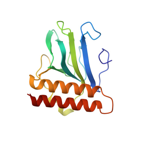
    The crystal structure of the arabinose-binding and dimerization domain of the Escherchia coli gene regulatory protein AraC was determined in the presence and absence of L-arabinose. The 1.5 angstrom structure of the arabinose-bound molecule shows that the protein adopts an unusual fold, binding sugar within a beta barrel and completely burying the arabinose with the amino-terminal arm of the protein. Dimer contacts in the presence of arabinose are mediated by an antiparallel coiled-coil. In the 2.8 angstrom structure of the uncomplexed protein, the amino-terminal arm is disordered, uncovering the sugar-binding pocket and allowing it to serve as an oligomerization interface. The ligand-gated oligomerization as seen in AraC provides the basis of a plausible mechanism for modulating the protein's DNA-looping properties.


  • Organizational Affiliation

    Department of Biophysics and Biophysical Chemistry, Johns Hopkins University School of Medicine, Baltimore, MD 21205, USA.


Macromolecules
Find similar proteins by:  (by identity cutoff)  |  3D Structure
Entity ID: 1
MoleculeChains Sequence LengthOrganismDetailsImage
ARABINOSE OPERON REGULATORY PROTEIN
A, B
164Escherichia coliMutation(s): 0 
UniProt
Find proteins for P0A9E0 (Escherichia coli (strain K12))
Explore P0A9E0 
Go to UniProtKB:  P0A9E0
Entity Groups  
Sequence Clusters30% Identity50% Identity70% Identity90% Identity95% Identity100% Identity
UniProt GroupP0A9E0
Sequence Annotations
Expand
  • Reference Sequence
Experimental Data & Validation

Experimental Data

  • Method: X-RAY DIFFRACTION
  • Resolution: 1.50 Å
  • R-Value Free:  0.232 (Depositor), 0.230 (DCC) 
  • R-Value Work:  0.179 (Depositor), 0.170 (DCC) 
  • R-Value Observed: 0.179 (Depositor) 
Space Group: P 1 21 1
Unit Cell:
Length ( Å )Angle ( ˚ )
a = 39.75α = 90
b = 93.84β = 95.62
c = 50.33γ = 90
Software Package:
Software NamePurpose
X-PLORmodel building
X-PLORrefinement
DENZOdata reduction
SCALEPACKdata scaling
X-PLORphasing

Structure Validation

View Full Validation Report


Ligand Structure Quality Assessment 

Created with Raphaël 2.3.0Worse 01 BetterLigand structure goodness of fit to experimental dataBest fitted ARAClick on this verticalbar to view details

Entry History 

Deposition Data

Revision History  (Full details and data files)

  • Version 1.0: 1997-05-15
    Type: Initial release
  • Version 1.1: 2008-03-03
    Changes: Version format compliance
  • Version 1.2: 2011-07-13
    Changes: Version format compliance
  • Version 1.3: 2020-07-29
    Type: Remediation
    Reason: Carbohydrate remediation
    Changes: Data collection, Derived calculations, Other, Structure summary
  • Version 1.4: 2024-02-14
    Changes: Data collection, Database references, Structure summary