2AR9

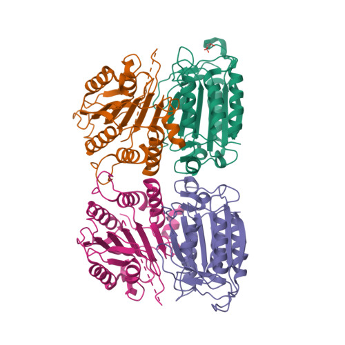
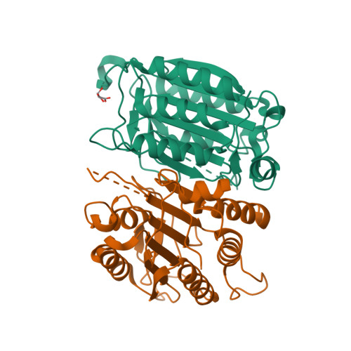
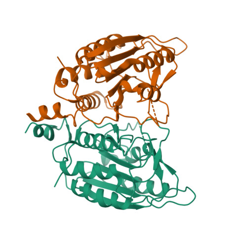
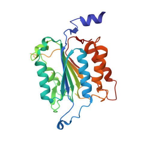
Crystal structure of a dimeric caspase-9


Experimental Data Snapshot

  • Method: X-RAY DIFFRACTION
  • Resolution: 2.80 Å
  • R-Value Free: 0.288 
  • R-Value Work: 0.237 
  • R-Value Observed: 0.240 

Starting Model: experimental
View more details

wwPDB Validation   3D Report Full Report


This is version 1.6 of the entry. See complete history


Literature

Engineering a Dimeric Caspase-9: A Re-Evaluation of the Induced Proximity Model for Caspase Activation

Chao, Y.Shiozaki, E.N.Srinivassula, S.M.Rigotti, D.J.Fairman, R.Shi, Y.

(2005) PLoS Biol 3: 1079-1087

  • DOI: https://doi.org/10.1371/journal.pbio.0030183
  • Primary Citation of Related Structures:  
    2AR9

  • PubMed Abstract: 

    Caspases are responsible for the execution of programmed cell death (apoptosis) and must undergo proteolytic activation, in response to apoptotic stimuli, to function. The mechanism of initiator caspase activation has been generalized by the induced proximity model, which is thought to drive dimerization-mediated activation of caspases. The initiator caspase, caspase-9, exists predominantly as a monomer in solution. To examine the induced proximity model, we engineered a constitutively dimeric caspase-9 by relieving steric hindrance at the dimer interface. Crystal structure of the engineered caspase-9 closely resembles that of the wild-type (WT) caspase-9, including all relevant structural details and the asymmetric nature of two monomers. Compared to the WT caspase-9, this engineered dimer exhibits a higher level of catalytic activity in vitro and induces more efficient cell death when expressed. However, the catalytic activity of the dimeric caspase-9 is only a small fraction of that for the Apaf-1-activated caspase-9. Furthermore, in contrast to the WT caspase-9, the activity of the dimeric caspase-9 can no longer be significantly enhanced in an Apaf-1-dependent manner. These findings suggest that dimerization of caspase-9 may be qualitatively different from its activation by Apaf-1, and in conjunction with other evidence, posit an induced conformation model for the activation of initiator caspases.


  • Organizational Affiliation

    Department of Molecular Biology, Lewis Thomas Laboratory, Princeton University, Princeton, New Jersey, USA.


Macromolecules
Find similar proteins by:  (by identity cutoff)  |  3D Structure
Entity ID: 1
MoleculeChains Sequence LengthOrganismDetailsImage
Caspase-9
A, B, C, D
278Homo sapiensMutation(s): 6 
EC: 3.4.22 (PDB Primary Data), 3.4.22.62 (UniProt)
UniProt & NIH Common Fund Data Resources
Find proteins for P55211 (Homo sapiens)
Explore P55211 
Go to UniProtKB:  P55211
PHAROS:  P55211
GTEx:  ENSG00000132906 
Entity Groups  
Sequence Clusters30% Identity50% Identity70% Identity90% Identity95% Identity100% Identity
UniProt GroupP55211
Sequence Annotations
Expand
  • Reference Sequence
Small Molecules
Ligands 1 Unique
IDChains Name / Formula / InChI Key2D Diagram3D Interactions
MLT
Query on MLT

Download Ideal Coordinates CCD File 
E [auth A]D-MALATE
C4 H6 O5
BJEPYKJPYRNKOW-UWTATZPHSA-N
Experimental Data & Validation

Experimental Data

  • Method: X-RAY DIFFRACTION
  • Resolution: 2.80 Å
  • R-Value Free: 0.288 
  • R-Value Work: 0.237 
  • R-Value Observed: 0.240 
  • Space Group: C 1 2 1
Unit Cell:
Length ( Å )Angle ( ˚ )
a = 144.698α = 90
b = 78.055β = 112.5
c = 125.963γ = 90
Software Package:
Software NamePurpose
DENZOdata reduction
SCALEPACKdata scaling
AMoREphasing
CNSrefinement

Structure Validation

View Full Validation Report


Entry History 

Deposition Data

Revision History  (Full details and data files)

  • Version 1.0: 2005-10-11
    Type: Initial release
  • Version 1.1: 2008-04-30
    Changes: Version format compliance
  • Version 1.2: 2011-07-13
    Changes: Version format compliance
  • Version 1.3: 2011-11-16
    Changes: Atomic model
  • Version 1.4: 2012-02-15
    Changes: Non-polymer description
  • Version 1.5: 2021-10-20
    Changes: Database references, Derived calculations
  • Version 1.6: 2023-08-23
    Changes: Data collection, Refinement description