2ANQ

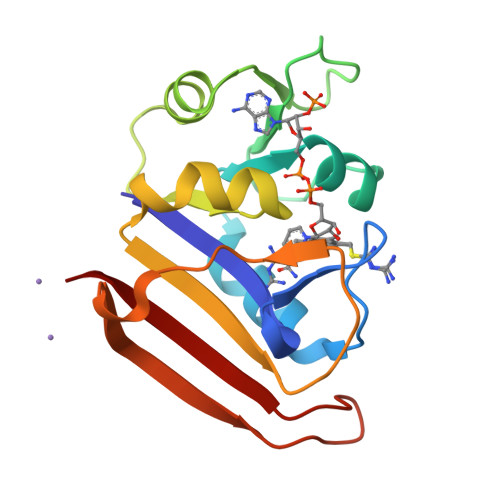
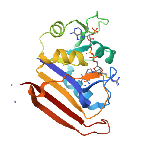
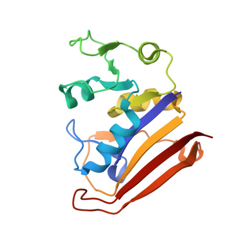
Crystal Structure of E.coli DHFR in complex with NADPH and the inhibitor compound 10a.


Experimental Data Snapshot

  • Method: X-RAY DIFFRACTION
  • Resolution: 2.13 Å
  • R-Value Free: 0.256 
  • R-Value Work: 0.233 
  • R-Value Observed: 0.235 

wwPDB Validation   3D Report Full Report


Ligand Structure Quality Assessment 


This is version 1.4 of the entry. See complete history


Literature

A 2.13 A Structure of E. coli Dihydrofolate Reductase Bound to a Novel Competitive Inhibitor Reveals a New Binding Surface Involving the M20 Loop Region

Summerfield, R.L.Daigle, D.M.Mayer, S.Mallik, D.Hughes, D.W.Jackson, S.G.Sulek, M.Organ, M.G.Brown, E.D.Junop, M.S.

(2006) J Med Chem 49: 6977-6986

  • DOI: https://doi.org/10.1021/jm060570v
  • Primary Citation of Related Structures:  
    2ANO, 2ANQ

  • PubMed Abstract: 

    Dihydrofolate reductase (DHFR) is a vital metabolic enzyme and thus a clinically prominent target in the design of antimetabolites. In this work, we identify 1,4-bis-{[N-(1-imino-1-guanidino-methyl)]sulfanylmethyl}-3,6-dimethyl-benzene (compound 1) as the correct structure of the previously reported DHFR inhibitor 1,4-bis-{(iminothioureidomethyl)aminomethyl}-3,6-dimethyl-benzene (compound 2). The fact that compound 1 has an uncharacteristic structure for DHFR inhibitors, and an affinity (KI of 11.5 nM) comparable to potent inhibitors such as methotrexate and trimethoprim, made this inhibitor of interest for further analysis. We have conducted a characterization of the primary interactions of compound 1 and DHFR using a combination of X-ray structure and SAR analysis. The crystal structure of E. coli DHFR in complex with compound 1 and NADPH reveals that one portion of this inhibitor exploits a unique binding surface, the M20 loop. The importance of this interface was further confirmed by SAR analysis and additional structural characterization.


  • Organizational Affiliation

    Atlantic Veterinary College, University of Prince Edward Island, 550 University Avenue, Charlottetown, Prince Edward Island C1A 4P3, Canada.


Macromolecules
Find similar proteins by:  (by identity cutoff)  |  3D Structure
Entity ID: 1
MoleculeChains Sequence LengthOrganismDetailsImage
Dihydrofolate reductase159Escherichia coliMutation(s): 1 
Gene Names: folAtmrA
EC: 1.5.1.3
UniProt
Find proteins for P0ABQ4 (Escherichia coli (strain K12))
Explore P0ABQ4 
Go to UniProtKB:  P0ABQ4
Entity Groups  
Sequence Clusters30% Identity50% Identity70% Identity90% Identity95% Identity100% Identity
UniProt GroupP0ABQ4
Sequence Annotations
Expand
  • Reference Sequence
Small Molecules
Binding Affinity Annotations 
IDSourceBinding Affinity
C1A BindingDB:  2ANQ Ki: 11.5 (nM) from 1 assay(s)
Kd: 13 (nM) from 1 assay(s)
IC50: 75 (nM) from 1 assay(s)
Experimental Data & Validation

Experimental Data

  • Method: X-RAY DIFFRACTION
  • Resolution: 2.13 Å
  • R-Value Free: 0.256 
  • R-Value Work: 0.233 
  • R-Value Observed: 0.235 
  • Space Group: P 21 21 21
Unit Cell:
Length ( Å )Angle ( ˚ )
a = 34α = 90
b = 44.72β = 90
c = 98.05γ = 90
Software Package:
Software NamePurpose
d*TREKdata processing
CNSrefinement
PDB_EXTRACTdata extraction
d*TREKdata reduction
d*TREKdata scaling
CNSphasing

Structure Validation

View Full Validation Report


Ligand Structure Quality Assessment 


Entry History 

Deposition Data

Revision History  (Full details and data files)

  • Version 1.0: 2006-07-25
    Type: Initial release
  • Version 1.1: 2008-04-30
    Changes: Version format compliance
  • Version 1.2: 2011-07-13
    Changes: Version format compliance
  • Version 1.3: 2021-10-20
    Changes: Database references, Derived calculations
  • Version 1.4: 2024-02-14
    Changes: Data collection