2AM1 | pdb_00002am1

sp protein ligand 1


Experimental Data Snapshot

  • Method: X-RAY DIFFRACTION
  • Resolution: 2.50 Å
  • R-Value Free: 
    0.290 (Depositor), 0.289 (DCC) 
  • R-Value Work: 
    0.238 (Depositor), 0.238 (DCC) 
  • R-Value Observed: 
    0.238 (Depositor) 

wwPDB Validation 3D Report Full Report

Validation slider image for 2AM1

Ligand Structure Quality Assessment 


This is version 1.4 of the entry. See complete history

Literature

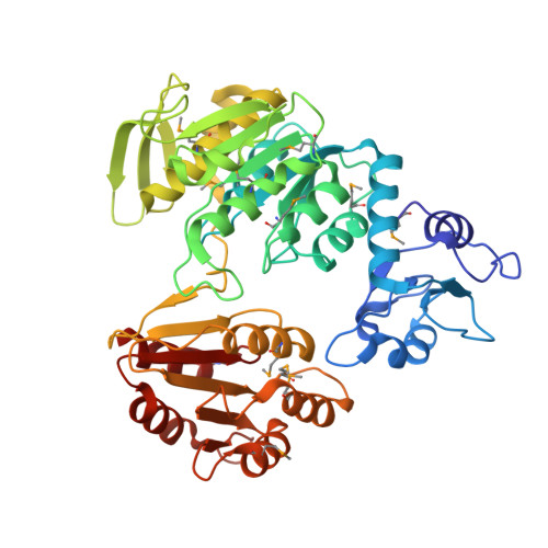
Structure of MurF from Streptococcus pneumoniae co-crystallized with a small molecule inhibitor exhibits interdomain closure

Longenecker, K.L.Stamper, G.F.Hajduk, P.J.Fry, E.H.Jakob, C.G.Harlan, J.E.Edalji, R.Bartley, D.M.Walter, K.A.Solomon, L.R.Holzman, T.F.Gu, Y.G.Lerner, C.G.Beutel, B.A.Stoll, V.S.

(2005) Protein Sci 14: 3039-3047

  • DOI: https://doi.org/10.1110/ps.051604805
  • Primary Citation Related Structures: 
    2AM1, 2AM2

  • PubMed Abstract: 

    In a broad genomics analysis to find novel protein targets for antibiotic discovery, MurF was identified as an essential gene product for Streptococcus pneumonia that catalyzes a critical reaction in the biosynthesis of the peptidoglycan in the formation of the cell wall. Lacking close relatives in mammalian biology, MurF presents attractive characteristics as a potential drug target. Initial screening of the Abbott small-molecule compound collection identified several compounds for further validation as pharmaceutical leads. Here we report the integrated efforts of NMR and X-ray crystallography, which reveal the multidomain structure of a MurF-inhibitor complex in a compact conformation that differs dramatically from related structures. The lead molecule is bound in the substrate-binding region and induces domain closure, suggestive of the domain arrangement for the as yet unobserved transition state conformation for MurF enzymes. The results form a basis for directed optimization of the compound lead by structure-based design to explore the suitability of MurF as a pharmaceutical target.


  • Organizational Affiliation
    • Department of Structural Biology, R46Y, Building AP10, 100 Abbott Park Road, Abbott Park, IL 60064, USA. Kenton.Longenecker@Abbott.com

Macromolecule Content 

  • Total Structure Weight: 51.34 kDa 
  • Atom Count: 3,675 
  • Modeled Residue Count: 454 
  • Deposited Residue Count: 454 
  • Unique protein chains: 1

Macromolecules

Find similar proteins by:|  3D Structure
Entity ID: 1
MoleculeChains  Sequence LengthOrganismDetailsImage
UDP-N-acetylmuramoylalanine-D-glutamyl-lysine-D-alanyl-D-alanine ligase, MurF protein454Streptococcus pneumoniae R6Mutation(s): 12 
Gene Names: murF
EC: 6.3.2.10
UniProt
Find proteins for Q8DNV6 (Streptococcus pneumoniae (strain ATCC BAA-255 / R6))
Explore Q8DNV6 
Go to UniProtKB:  Q8DNV6
Entity Groups
Sequence Clusters30% Identity50% Identity70% Identity90% Identity95% Identity100% Identity
UniProt GroupQ8DNV6
Sequence Annotations
Expand
Reference Sequence

Small Molecules

Ligands 2 Unique
IDChains Name / Formula / InChI Key2D Diagram3D Interactions
1LG

Query on 1LG


Download:Ideal Coordinates CCD File
B [auth A]2,4-DICHLORO-N-(3-CYANO-4,5,6,7-TETRAHYDRO-BENZOTHIOPHEN-2YL)-5-(MORPHOLINE-4-SULFONYL)-BENZAMIDE
C20 H19 Cl2 N3 O4 S2
JMCVGASJAJMFPN-UHFFFAOYSA-N
GOL

Query on GOL


Download:Ideal Coordinates CCD File
C [auth A]GLYCEROL
C3 H8 O3
PEDCQBHIVMGVHV-UHFFFAOYSA-N
Modified Residues  1 Unique
IDChains TypeFormula2D DiagramParent
MSE
Query on MSE
A
L-PEPTIDE LINKINGC5 H11 N O2 SeMET
Binding Affinity Annotations 
IDSourceBinding Affinity
1LG BindingDB:  2AM1 IC50: 1000 (nM) from 1 assay(s)

Experimental Data & Validation

Experimental Data

  • Method: X-RAY DIFFRACTION
  • Resolution: 2.50 Å
  • R-Value Free:  0.290 (Depositor), 0.289 (DCC) 
  • R-Value Work:  0.238 (Depositor), 0.238 (DCC) 
  • R-Value Observed: 0.238 (Depositor) 
Space Group: P 61 2 2
Unit Cell:
Length ( Å )Angle ( ˚ )
a = 116.271α = 90
b = 116.271β = 90
c = 161.389γ = 120
Software Package:
Software NamePurpose
DENZOdata reduction
SCALEPACKdata scaling
CNSrefinement
PDB_EXTRACTdata extraction
HKL-2000data reduction
CCP4data scaling
SOLVEphasing

Structure Validation

View Full Validation Report


Ligand Structure Quality Assessment 


Entry History 

Deposition Data

Revision History  (Full details and data files)

  • Version 1.0: 2006-01-24
    Type: Initial release
  • Version 1.1: 2008-04-30
    Changes: Version format compliance
  • Version 1.2: 2011-07-13
    Changes: Source and taxonomy, Version format compliance
  • Version 1.3: 2017-10-11
    Changes: Refinement description
  • Version 1.4: 2024-10-30
    Changes: Data collection, Database references, Derived calculations, Structure summary