2AIQ | pdb_00002aiq

Crystal structure of benzamidine-inhibited protein C activator from the venom of copperhead snake Agkistrodon contortrix contortrix


Experimental Data Snapshot

  • Method: X-RAY DIFFRACTION
  • Resolution: 1.54 Å
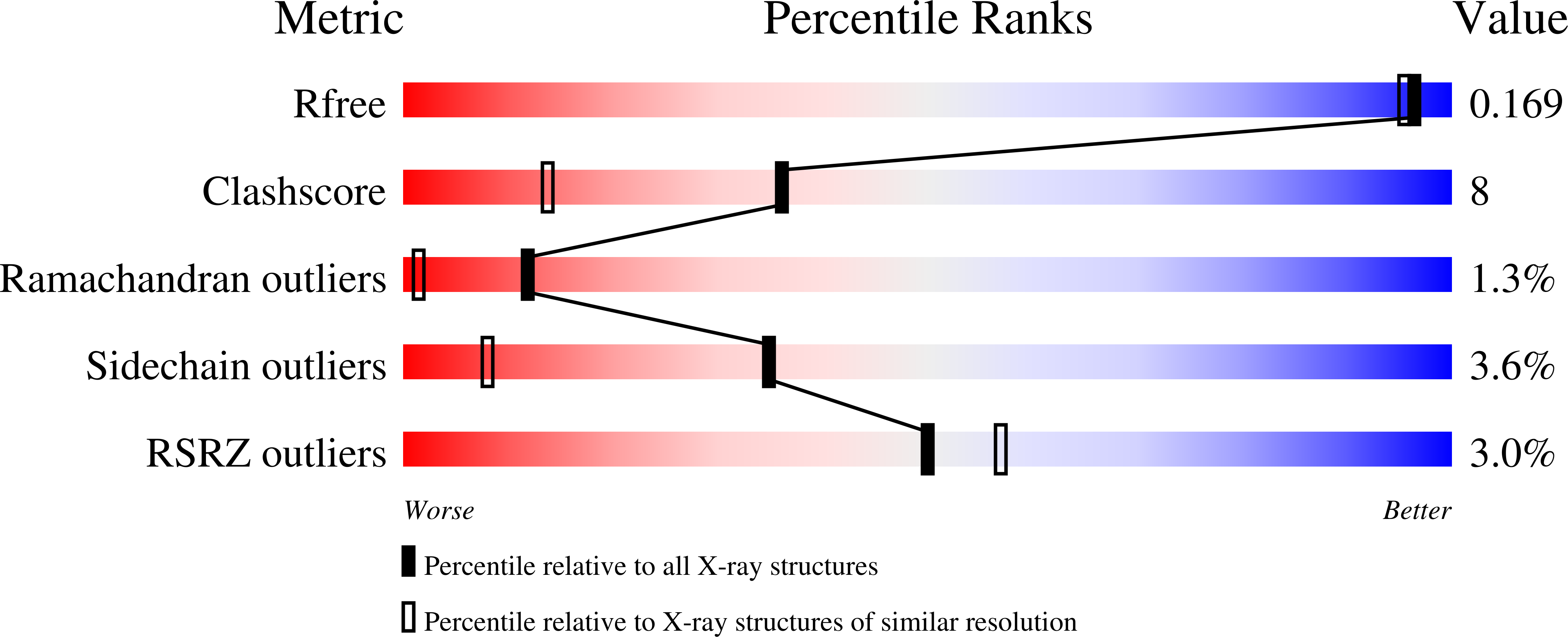
  • R-Value Free: 
    0.191 (Depositor), 0.170 (DCC) 
  • R-Value Work: 
    0.168 (Depositor), 0.170 (DCC) 
  • R-Value Observed: 
    0.169 (Depositor) 

Starting Model: experimental
View more details

wwPDB Validation   3D Report Full Report


Ligand Structure Quality Assessment 

Created with Raphaël 2.3.0Worse 01 BetterLigand structure goodness of fit to experimental dataBest fitted NAGClick on this verticalbar to view details

This is version 2.2 of the entry. See complete history


Literature

Thrombomodulin-independent Activation of Protein C and Specificity of Hemostatically Active Snake Venom Serine Proteinases: CRYSTAL STRUCTURES OF NATIVE AND INHIBITED AGKISTRODON CONTORTRIX CONTORTRIX PROTEIN C ACTIVATOR.

Murakami, M.T.Arni, R.K.

(2005) J Biological Chem 280: 39309-39315

  • DOI: https://doi.org/10.1074/jbc.M508502200
  • Primary Citation of Related Structures:  
    2AIP, 2AIQ

  • PubMed Abstract: 

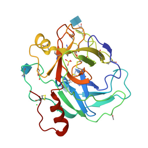
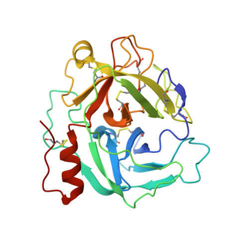
    Protein C activation initiated by the thrombin-thrombomodulin complex forms the major physiological anticoagulant pathway. Agkistrodon contortrix contortrix protein C activator, a glycosylated single-chain serine proteinase, activates protein C without relying on thrombomodulin. The crystal structures of native and inhibited Agkistrodon contortrix contortrix protein C activator determined at 1.65 and 1.54 A resolutions, respectively, indicate the pivotal roles played by the positively charged belt and the strategic positioning of the three carbohydrate moieties surrounding the catalytic site in protein C recognition, binding, and activation. Structural changes in the benzamidine-inhibited enzyme suggest a probable function in allosteric regulation for the anion-binding site located in the C-terminal extension, which is fully conserved in snake venom serine proteinases, that preferentially binds Cl(1-) instead of SO(4)(2-).


  • Organizational Affiliation

    Biochemistry and Structural Biology Group, Department of Physics, IBILCE/UNESP, São José do Rio Preto, SP 15054-000, Brazil.


Macromolecules
Find similar proteins by:  (by identity cutoff)  |  3D Structure
Entity ID: 1
MoleculeChains Sequence LengthOrganismDetailsImage
Protein C activator231Agkistrodon contortrix contortrixMutation(s): 0 
EC: 3.4.21.74 (PDB Primary Data), 3.4.21 (UniProt)
UniProt
Find proteins for P09872 (Agkistrodon contortrix contortrix)
Explore P09872 
Go to UniProtKB:  P09872
Entity Groups  
Sequence Clusters30% Identity50% Identity70% Identity90% Identity95% Identity100% Identity
UniProt GroupP09872
Glycosylation
Glycosylation Sites: 3
Sequence Annotations
Expand
  • Reference Sequence
Small Molecules
Ligands 6 Unique
IDChains Name / Formula / InChI Key2D Diagram3D Interactions
NAG
Query on NAG

Download Ideal Coordinates CCD File 
B [auth A],
C [auth A],
D [auth A]
2-acetamido-2-deoxy-beta-D-glucopyranose
C8 H15 N O6
OVRNDRQMDRJTHS-FMDGEEDCSA-N
BEN
Query on BEN

Download Ideal Coordinates CCD File 
H [auth A]BENZAMIDINE
C7 H8 N2
PXXJHWLDUBFPOL-UHFFFAOYSA-N
SO4
Query on SO4

Download Ideal Coordinates CCD File 
E [auth A]SULFATE ION
O4 S
QAOWNCQODCNURD-UHFFFAOYSA-L
GOL
Query on GOL

Download Ideal Coordinates CCD File 
I [auth A]GLYCEROL
C3 H8 O3
PEDCQBHIVMGVHV-UHFFFAOYSA-N
ACT
Query on ACT

Download Ideal Coordinates CCD File 
F [auth A]ACETATE ION
C2 H3 O2
QTBSBXVTEAMEQO-UHFFFAOYSA-M
CL
Query on CL

Download Ideal Coordinates CCD File 
G [auth A]CHLORIDE ION
Cl
VEXZGXHMUGYJMC-UHFFFAOYSA-M
Experimental Data & Validation

Experimental Data

  • Method: X-RAY DIFFRACTION
  • Resolution: 1.54 Å
  • R-Value Free:  0.191 (Depositor), 0.170 (DCC) 
  • R-Value Work:  0.168 (Depositor), 0.170 (DCC) 
  • R-Value Observed: 0.169 (Depositor) 
Space Group: C 1 2 1
Unit Cell:
Length ( Å )Angle ( ˚ )
a = 80.516α = 90
b = 63.463β = 99.85
c = 48.216γ = 90
Software Package:
Software NamePurpose
REFMACrefinement
DENZOdata reduction
SCALEPACKdata scaling
AMoREphasing

Structure Validation

View Full Validation Report


Ligand Structure Quality Assessment 

Created with Raphaël 2.3.0Worse 01 BetterLigand structure goodness of fit to experimental dataBest fitted NAGClick on this verticalbar to view details

Entry History 

Deposition Data

Revision History  (Full details and data files)

  • Version 1.0: 2005-09-06
    Type: Initial release
  • Version 1.1: 2008-04-30
    Changes: Version format compliance
  • Version 1.2: 2011-07-13
    Changes: Non-polymer description, Version format compliance
  • Version 2.0: 2020-07-29
    Type: Remediation
    Reason: Carbohydrate remediation
    Changes: Advisory, Atomic model, Data collection, Derived calculations, Non-polymer description, Structure summary
  • Version 2.1: 2023-08-23
    Changes: Data collection, Database references, Refinement description, Structure summary
  • Version 2.2: 2024-11-06
    Changes: Structure summary