2AES | pdb_00002aes

Crystal Structure of Human M340H-Beta1,4-Galactosyltransferase-I (M340H-B4Gal-T1) in Complex with GlcNAc-beta1,2-Man-alpha1,3-Man-beta-OR


Experimental Data Snapshot

  • Method: X-RAY DIFFRACTION
  • Resolution: 2.00 Å
  • R-Value Free: 
    0.232 (Depositor) 
  • R-Value Work: 
    0.204 (Depositor), 0.190 (DCC) 
  • R-Value Observed: 
    0.204 (Depositor) 

Starting Model: experimental
View more details

wwPDB Validation   3D Report Full Report


Ligand Structure Quality Assessment 

Created with Raphaël 2.3.0Worse 01 BetterLigand structure goodness of fit to experimental dataBest fitted UDHClick on this verticalbar to view details

This is version 2.3 of the entry. See complete history


Literature

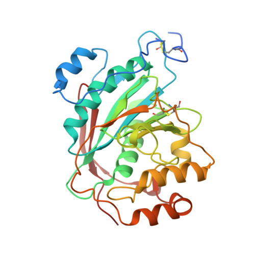
Oligosaccharide Preferences of beta1,4-Galactosyltransferase-I: Crystal Structures of Met340His Mutant of Human beta1,4-Galactosyltransferase-I with a Pentasaccharide and Trisaccharides of the N-Glycan Moiety

Ramasamy, V.Ramakrishnan, B.Boeggeman, E.Ratner, D.M.Seeberger, P.H.Qasba, P.K.

(2005) J Mol Biology 353: 53-67

  • DOI: https://doi.org/10.1016/j.jmb.2005.07.050
  • Primary Citation of Related Structures:  
    2AE7, 2AEC, 2AES, 2AGD, 2AH9

  • PubMed Abstract: 

    beta-1,4-Galactosyltransferase-I (beta4Gal-T1) transfers galactose from UDP-galactose to N-acetylglucosamine (GlcNAc) residues of the branched N-linked oligosaccharide chains of glycoproteins. In an N-linked biantennary oligosaccharide chain, one antenna is attached to the 3-hydroxyl-(1,3-arm), and the other to the 6-hydroxyl-(1,6-arm) group of mannose, which is beta-1,4-linked to an N-linked chitobiose, attached to the aspargine residue of a protein. For a better understanding of the branch specificity of beta4Gal-T1 towards the GlcNAc residues of N-glycans, we have carried out kinetic and crystallographic studies with the wild-type human beta4Gal-T1 (h-beta4Gal-T1) and the mutant Met340His-beta4Gal-T1 (h-M340H-beta4Gal-T1) in complex with a GlcNAc-containing pentasaccharide and several GlcNAc-containing trisaccharides present in N-glycans. The oligosaccharides used were: pentasaccharide GlcNAcbeta1,2-Manalpha1,6 (GlcNAcbeta1,2-Manalpha1,3)Man; the 1,6-arm trisaccharide, GlcNAcbeta1,2-Manalpha1,6-Manbeta-OR (1,2-1,6-arm); the 1,3-arm trisaccharides, GlcNAcbeta1,2-Manalpha1,3-Manbeta-OR (1,2-1,3-arm) and GlcNAcbeta1,4-Manalpha1,3-Manbeta-OR (1,4-1,3-arm); and the trisaccharide GlcNAcbeta1,4-GlcNAcbeta1,4-GlcNAc (chitotriose). With the wild-type h-beta4Gal-T1, the K(m) of 1,2-1,6-arm is approximately tenfold lower than for 1,2-1,3-arm and 1,4-1,3-arm, and 22-fold lower than for chitotriose. Crystal structures of h-M340H-beta4Gal-T1 in complex with the pentasaccharide and various trisaccharides at 1.9-2.0A resolution showed that beta4Gal-T1 is in a closed conformation with the oligosaccharide bound to the enzyme, and the 1,2-1,6-arm trisaccharide makes the maximum number of interactions with the enzyme, which is in concurrence with the lowest K(m) for the trisaccharide. Present studies suggest that beta4Gal-T1 interacts preferentially with the 1,2-1,6-arm trisaccharide rather than with the 1,2-1,3-arm or 1,4-1,3-arm of a bi- or tri-antennary oligosaccharide chain of N-glycan.


  • Organizational Affiliation

    Laboratory of Experimental and Computational Biology, Center for Cancer Research, National Cancer Institute at Frederick, Frederick, MD 21702, USA.


Macromolecules
Find similar proteins by:  (by identity cutoff)  |  3D Structure
Entity ID: 1
MoleculeChains Sequence LengthOrganismDetailsImage
Beta-1,4-galactosyltransferase 1
A, B, C
287Homo sapiensMutation(s): 3 
Gene Names: B4GALT1GGTB2
EC: 2.4.1.90 (PDB Primary Data), 2.4.1 (UniProt), 2.4.1.22 (UniProt), 2.4.1.275 (UniProt), 2.4.1.38 (UniProt)
UniProt & NIH Common Fund Data Resources
Find proteins for P15291 (Homo sapiens)
Explore P15291 
Go to UniProtKB:  P15291
PHAROS:  P15291
GTEx:  ENSG00000086062 
Entity Groups  
Sequence Clusters30% Identity50% Identity70% Identity90% Identity95% Identity100% Identity
UniProt GroupP15291
Sequence Annotations
Expand
  • Reference Sequence
Oligosaccharides

Help

Entity ID: 2
MoleculeChains Length2D Diagram Glycosylation3D Interactions
2-acetamido-2-deoxy-beta-D-glucopyranose-(1-2)-alpha-D-mannopyranose-(1-3)-beta-D-mannopyranose
D, E, F
3N/A
Glycosylation Resources
GlyTouCan:  G71817IG
GlyCosmos:  G71817IG
GlyGen:  G71817IG
Small Molecules
Ligands 6 Unique
IDChains Name / Formula / InChI Key2D Diagram3D Interactions
UDH
Query on UDH

Download Ideal Coordinates CCD File 
FA [auth C],
L [auth A],
W [auth B]
6-AMINOHEXYL-URIDINE-C1,5'-DIPHOSPHATE
C15 H27 N3 O12 P2
MLWJBKPFDKRHBM-FMKGYKFTSA-N
MES
Query on MES

Download Ideal Coordinates CCD File 
HA [auth C]2-(N-MORPHOLINO)-ETHANESULFONIC ACID
C6 H13 N O4 S
SXGZJKUKBWWHRA-UHFFFAOYSA-N
SO4
Query on SO4

Download Ideal Coordinates CCD File 
CA [auth C]
DA [auth C]
EA [auth C]
H [auth A]
I [auth A]
SULFATE ION
O4 S
QAOWNCQODCNURD-UHFFFAOYSA-L
GOL
Query on GOL

Download Ideal Coordinates CCD File 
AA [auth B]
IA [auth C]
M [auth A]
N [auth A]
O [auth A]
GLYCEROL
C3 H8 O3
PEDCQBHIVMGVHV-UHFFFAOYSA-N
DIO
Query on DIO

Download Ideal Coordinates CCD File 
GA [auth C]1,4-DIETHYLENE DIOXIDE
C4 H8 O2
RYHBNJHYFVUHQT-UHFFFAOYSA-N
MN
Query on MN

Download Ideal Coordinates CCD File 
BA [auth C],
G [auth A],
P [auth B]
MANGANESE (II) ION
Mn
WAEMQWOKJMHJLA-UHFFFAOYSA-N
Experimental Data & Validation

Experimental Data

  • Method: X-RAY DIFFRACTION
  • Resolution: 2.00 Å
  • R-Value Free:  0.232 (Depositor) 
  • R-Value Work:  0.204 (Depositor), 0.190 (DCC) 
  • R-Value Observed: 0.204 (Depositor) 
Space Group: C 2 2 21
Unit Cell:
Length ( Å )Angle ( ˚ )
a = 107.159α = 90
b = 192.989β = 90
c = 143.428γ = 90
Software Package:
Software NamePurpose
HKL-2000data collection
SCALEPACKdata scaling
AMoREphasing
CNSrefinement
HKL-2000data reduction

Structure Validation

View Full Validation Report


Ligand Structure Quality Assessment 

Created with Raphaël 2.3.0Worse 01 BetterLigand structure goodness of fit to experimental dataBest fitted UDHClick on this verticalbar to view details

Entry History 

Deposition Data

Revision History  (Full details and data files)

  • Version 1.0: 2005-10-04
    Type: Initial release
  • Version 1.1: 2008-04-30
    Changes: Version format compliance
  • Version 1.2: 2011-07-13
    Changes: Non-polymer description, Version format compliance
  • Version 2.0: 2020-07-29
    Type: Remediation
    Reason: Carbohydrate remediation
    Changes: Atomic model, Data collection, Derived calculations, Structure summary
  • Version 2.1: 2021-10-20
    Changes: Database references, Structure summary
  • Version 2.2: 2023-08-23
    Changes: Data collection, Refinement description
  • Version 2.3: 2024-11-13
    Changes: Structure summary