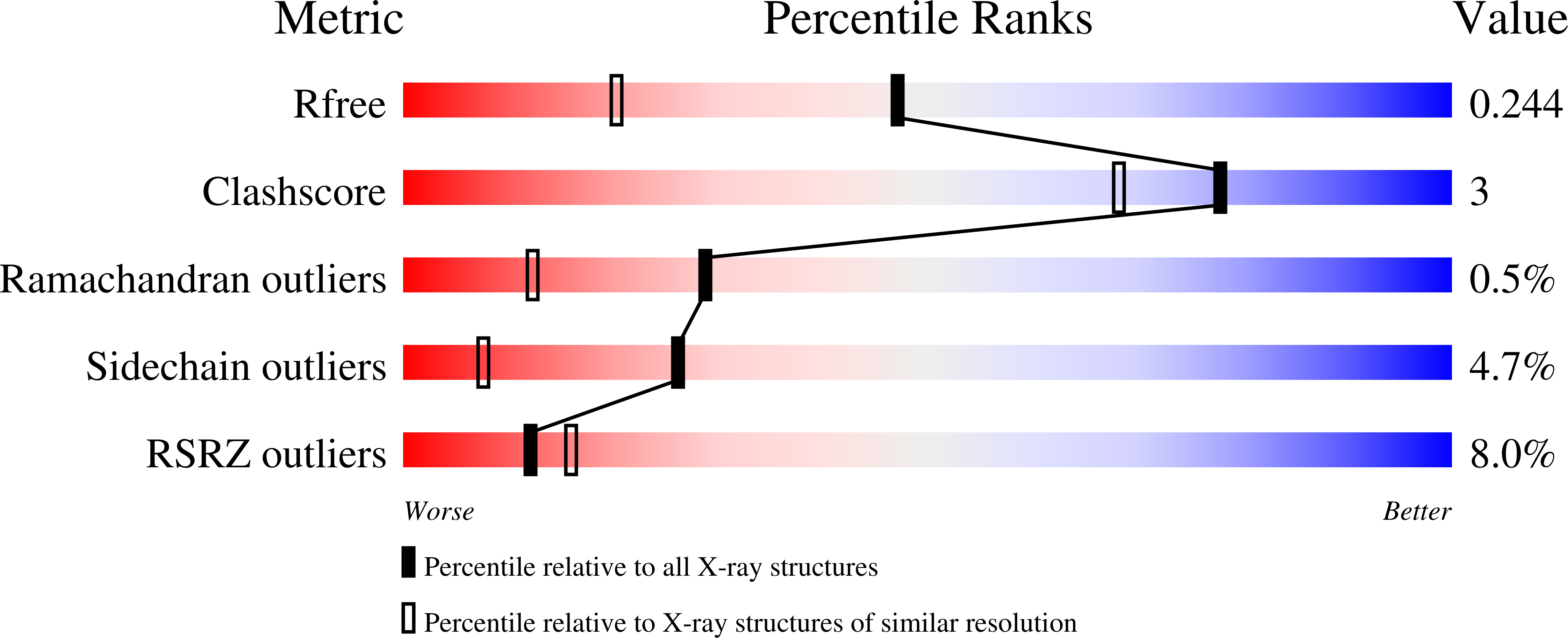
Structures of the carbohydrate recognition domain of Ca2+-independent cargo receptors Emp46p and Emp47p.
Satoh, T., Sato, K., Kanoh, A., Yamashita, K., Yamada, Y., Igarashi, N., Kato, R., Nakano, A., Wakatsuki, S.(2006) J Biological Chem 281: 10410-10419
- PubMed: 16439369
- DOI: https://doi.org/10.1074/jbc.M512258200
- Primary Citation of Related Structures:
2A6V, 2A6W, 2A6X, 2A6Y, 2A6Z, 2A70, 2A71 - PubMed Abstract:
Emp46p and Emp47p are type I membrane proteins, which cycle between the endoplasmic reticulum (ER) and the Golgi apparatus by vesicles coated with coat protein complexes I and II (COPI and COPII). They are considered to function as cargo receptors for exporting N-linked glycoproteins from the ER. We have determined crystal structures of the carbohydrate recognition domains (CRDs) of Emp46p and Emp47p of Saccharomyces cerevisiae, in the absence and presence of metal ions. Both proteins fold as a beta-sandwich, and resemble that of the mammalian ortholog, p58/ERGIC-53. However, the nature of metal binding is distinct from that of Ca(2+)-dependent p58/ERGIC-53. Interestingly, the CRD of Emp46p does not bind Ca(2+) ion but instead binds K(+) ion at the edge of a concave beta-sheet whose position is distinct from the corresponding site of the Ca(2+) ion in p58/ERGIC-53. Binding of K(+) ion to Emp46p appears essential for transport of a subset of glycoproteins because the Y131F mutant of Emp46p, which cannot bind K(+) ion fails to rescue the transport in disruptants of EMP46 and EMP47 genes. In contrast the CRD of Emp47p binds no metal ions at all. Furthermore, the CRD of Emp46p binds to glycoproteins carrying high mannosetype glycans and the is promoted by binding not the addition of Ca(2+) or K(+) ion in These results suggest that Emp46p can be regarded as a Ca(2+)-independent intracellular lectin at the ER exit sites.
Organizational Affiliation:
Structural Biology Research Center, Photon Factory, Institute of Materials Structure Science, High Energy Accelerator Research Organization (KEK), Tsukuba, Ibaraki 305-0801, Japan.


