2A5X | pdb_00002a5x

Crystal Structure of a Cross-linked Actin Dimer


Experimental Data Snapshot

  • Method: X-RAY DIFFRACTION
  • Resolution: 2.49 Å
  • R-Value Free: 
    0.250 (Depositor), 0.246 (DCC) 
  • R-Value Work: 
    0.194 (Depositor), 0.195 (DCC) 
  • R-Value Observed: 
    0.197 (Depositor) 

Starting Model: experimental
View more details

wwPDB Validation 3D Report Full Report

Validation slider image for 2A5X

Ligand Structure Quality Assessment 


This is version 1.4 of the entry. See complete history

Literature

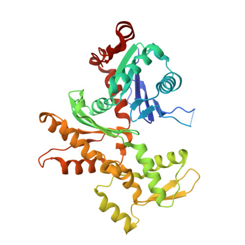
The crystal structure of a cross-linked actin dimer suggests a detailed molecular interface in F-actin

Kudryashov, D.S.Sawaya, M.R.Adisetiyo, H.Norcross, T.Hegyi, G.Reisler, E.Yeates, T.O.

(2005) Proc Natl Acad Sci U S A 102: 13105-13110

  • DOI: https://doi.org/10.1073/pnas.0506429102
  • Primary Citation Related Structures: 
    2A5X

  • PubMed Abstract: 

    The 2.5-A resolution crystal structure is reported for an actin dimer, composed of two protomers cross-linked along the longitudinal (or vertical) direction of the F-actin filament. The crystal structure provides an atomic resolution view of a molecular interface between actin protomers, which we argue represents a near-native interaction in the F-actin filament. The interaction involves subdomains 3 and 4 from distinct protomers. The atomic positions in the interface visualized differ by 5-10 A from those suggested by previous models of F-actin. Such differences fall within the range of uncertainties allowed by the fiber diffraction and electron microscopy methods on which previous models have been based. In the crystal, the translational arrangement of protomers lacks the slow twist found in native filaments. A plausible model of F-actin can be constructed by reintroducing the known filament twist, without disturbing significantly the interface observed in the actin dimer crystal.


  • Organizational Affiliation
    • Department of Chemistry and Biochemistry and Molecular Biology Institute, University of California, Los Angeles, CA 90095, USA.

Macromolecule Content 

  • Total Structure Weight: 43.29 kDa 
  • Atom Count: 2,812 
  • Modeled Residue Count: 342 
  • Deposited Residue Count: 375 
  • Unique protein chains: 1

Macromolecules

Find similar proteins by:|  3D Structure
Entity ID: 1
MoleculeChains  Sequence LengthOrganismDetailsImage
Actin, alpha skeletal muscle375Oryctolagus cuniculusMutation(s): 0 
EC: 3.6.4
UniProt
Find proteins for P68135 (Oryctolagus cuniculus)
Explore P68135 
Go to UniProtKB:  P68135
Entity Groups
Sequence Clusters30% Identity50% Identity70% Identity90% Identity95% Identity100% Identity
UniProt GroupP68135
Sequence Annotations
Expand
Reference Sequence

Small Molecules

Ligands 5 Unique
IDChains Name / Formula / InChI Key2D Diagram3D Interactions
ANP

Query on ANP


Download:Ideal Coordinates CCD File
D [auth A]PHOSPHOAMINOPHOSPHONIC ACID-ADENYLATE ESTER
C10 H17 N6 O12 P3
PVKSNHVPLWYQGJ-KQYNXXCUSA-N
LAR

Query on LAR


Download:Ideal Coordinates CCD File
E [auth A]LATRUNCULIN A
C22 H31 N O5 S
DDVBPZROPPMBLW-IZGXTMSKSA-N
NSB

Query on NSB


Download:Ideal Coordinates CCD File
F [auth A]N,N,N-TRIMETHYL-3-SULFOPROPAN-1-AMINIUM
C6 H16 N O3 S
WFJHXXPYPMNRPK-UHFFFAOYSA-O
MPD

Query on MPD


Download:Ideal Coordinates CCD File
G [auth A],
H [auth A]
(4S)-2-METHYL-2,4-PENTANEDIOL
C6 H14 O2
SVTBMSDMJJWYQN-YFKPBYRVSA-N
CA

Query on CA


Download:Ideal Coordinates CCD File
B [auth A],
C [auth A]
CALCIUM ION
Ca
BHPQYMZQTOCNFJ-UHFFFAOYSA-N

Experimental Data & Validation

Experimental Data

  • Method: X-RAY DIFFRACTION
  • Resolution: 2.49 Å
  • R-Value Free:  0.250 (Depositor), 0.246 (DCC) 
  • R-Value Work:  0.194 (Depositor), 0.195 (DCC) 
  • R-Value Observed: 0.197 (Depositor) 
Space Group: C 1 2 1
Unit Cell:
Length ( Å )Angle ( ˚ )
a = 207.377α = 90
b = 54.372β = 98.62
c = 36.2γ = 90
Software Package:
Software NamePurpose
REFMACrefinement
Blu-Icedata collection
SCALEPACKdata scaling
EPMRphasing

Structure Validation

View Full Validation Report


Ligand Structure Quality Assessment 


Entry History 

Deposition Data

Revision History  (Full details and data files)

  • Version 1.0: 2005-08-23
    Type: Initial release
  • Version 1.1: 2008-04-30
    Changes: Version format compliance
  • Version 1.2: 2011-07-13
    Changes: Advisory, Version format compliance
  • Version 1.3: 2017-10-11
    Changes: Refinement description
  • Version 1.4: 2023-08-23
    Changes: Advisory, Data collection, Database references, Derived calculations, Refinement description