2A3H | pdb_00002a3h

CELLOBIOSE COMPLEX OF THE ENDOGLUCANASE CEL5A FROM BACILLUS AGARADHERANS AT 2.0 A RESOLUTION


Experimental Data Snapshot

  • Method: X-RAY DIFFRACTION
  • Resolution: 2.00 Å
  • R-Value Free: 
    0.190 (Depositor) 
  • R-Value Work: 
    0.137 (Depositor), 0.133 (DCC) 

wwPDB Validation 3D Report Full Report

Validation slider image for 2A3H

This is version 2.1 of the entry. See complete history

Literature

Structure of the Bacillus agaradherans family 5 endoglucanase at 1.6 A and its cellobiose complex at 2.0 A resolution

Davies, G.J.Dauter, M.Brzozowski, A.M.Bjornvad, M.E.Andersen, K.V.Schulein, M.

(1998) Biochemistry 37: 1926-1932

  • DOI: https://doi.org/10.1021/bi972162m
  • Primary Citation Related Structures: 
    1A3H, 2A3H

  • PubMed Abstract: 

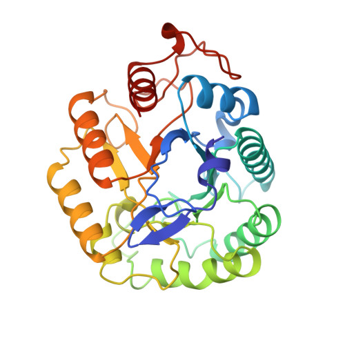
    The enzymatic degradation of cellulose, by cellulases, is not only industrially important in the food, paper, and textile industries but also a potentially useful method for the environmentally friendly recycling of municipal waste. An understanding of the structural and mechanistic requirements for the hydrolysis of the beta-1,4 glycosidic bonds of cellulose is an essential prerequisite for beneficial engineering of cellulases for these processes. Cellulases have been classified into 13 of the 62 glycoside hydrolase families [Henrissat, B., and Bairoch, A. (1996) Biochem J. 316, 695-696]. The structure of the catalytic core of the family 5 endoglucanase, Ce15A, from the alkalophilic Bacillus agaradherans has been solved by multiple isomorphous replacement at 1.6 A resolution. Ce15A has the (alpha/beta)8 barrel structure and signature structural features typical of the grouping of glycoside hydrolase families known as clan GH-A, with the catalytic acid/base Glu 139 and nucleophile Glu 228 on barrel strands beta 4 and beta 7 as expected. In addition to the native enzyme, the 2.0 A resolution structure of the cellobiose-bound form of the enzyme has also been determined. Cellobiose binds preferentially in the -2 and -3 subsites of the enzyme. Kinetic studies on the isolated catalytic core domain of Ce15A, using a series of reduced cellodextrins as substrates, suggest approximately five to six binding sites, consistent with the shape and size of the cleft observed by crystallography.


  • Organizational Affiliation
    • Department of Chemistry, University of York, Heslington, England. davies@yorvic.york.ac.uk

Macromolecule Content 

  • Total Structure Weight: 34 kDa 
  • Atom Count: 2,813 
  • Modeled Residue Count: 300 
  • Deposited Residue Count: 300 
  • Unique protein chains: 1

Macromolecules

Find similar proteins by:|  3D Structure
Entity ID: 1
MoleculeChains  Sequence LengthOrganismDetailsImage
ENDOGLUCANASE300Salipaludibacillus agaradhaerensMutation(s): 0 
EC: 3.2.1.4
UniProt
Find proteins for O85465 (Salipaludibacillus agaradhaerens)
Explore O85465 
Go to UniProtKB:  O85465
Entity Groups
Sequence Clusters30% Identity50% Identity70% Identity90% Identity95% Identity100% Identity
UniProt GroupO85465
Sequence Annotations
Expand
Reference Sequence

Oligosaccharides

Help  
Entity ID: 2
MoleculeChains Length2D Diagram GlycosylationD Interactions
beta-D-glucopyranose-(1-4)-beta-D-glucopyranose
B
2N/A
Glycosylation Resources
GlyTouCan: G84824ZO
GlyCosmos: G84824ZO

Biologically Interesting Molecules (External Reference) 

1 Unique

Experimental Data & Validation

Experimental Data

  • Method: X-RAY DIFFRACTION
  • Resolution: 2.00 Å
  • R-Value Free:  0.190 (Depositor) 
  • R-Value Work:  0.137 (Depositor), 0.133 (DCC) 
Space Group: P 21 21 21
Unit Cell:
Length ( Å )Angle ( ˚ )
a = 54.71α = 90
b = 69.57β = 90
c = 77.04γ = 90
Software Package:
Software NamePurpose
CCP4model building
REFMACrefinement
DENZOdata reduction
SCALEPACKdata scaling
CCP4phasing

Structure Validation

View Full Validation Report


Entry History 

Deposition Data

Revision History  (Full details and data files)

  • Version 1.0: 1999-03-16
    Type: Initial release
  • Version 1.1: 2008-03-03
    Changes: Version format compliance
  • Version 1.2: 2011-07-13
    Changes: Version format compliance
  • Version 2.0: 2020-07-29
    Type: Remediation
    Reason: Carbohydrate remediation
    Changes: Atomic model, Data collection, Derived calculations, Non-polymer description, Other, Structure summary
  • Version 2.1: 2024-02-14
    Changes: Data collection, Database references, Structure summary