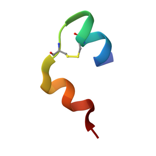
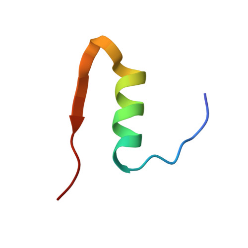
The structure of T6 bovine insulin.
Smith, G.D., Pangborn, W.A., Blessing, R.H.(2005) Acta Crystallogr D Biol Crystallogr 61: 1476-1482
- PubMed: 16239724
- DOI: https://doi.org/10.1107/S0907444905025771
- Primary Citation of Related Structures:
2A3G - PubMed Abstract:
Porcine insulin differs in sequence from bovine insulin at residues A8 (Thr in porcine-->Ala in bovine) and A10 (Ile in porcine-->Val in bovine). The structure of T6 hexameric bovine insulin has been determined to 2.25 A resolution at room temperature and refined to a residual of 0.162. The structure of the independent dimer is nearly identical to the T6 porcine insulin dimer: the mean displacement of all backbone atoms is 0.16 A, with the largest displacements occurring at AlaB30. Each of two independent zinc ions is octahedrally coordinated by three HisB10 side chains and three water molecules. As has been observed in both human and porcine insulin, the GluB13 side chains are directed towards the center of the hexamer, where a short contact of 2.57 A occurs between two independent carboxyl O atoms, again suggesting the presence of a centered hydrogen bond. No significant displacements of backbone atoms or changes in conformation are observed at A8 or A10. Since there are no interhexamer hydrogen-bonded contacts involving A8 in either porcine or bovine insulin, the change in the identity of this residue appears to have little or no effect upon the packing of the hexamers in the unit cell. In contrast, the side chains of the three A10 residues in one trimer make van der Waals contacts with the A10 side chains in a translationally related hexamer. As a consequence of the loss of the C(delta1) atom from the isoleucine residue in porcine insulin to produce valine in bovine insulin, there is a 0.36 A decrease in the distance between independent pairs of C(beta) atoms and a 0.24 A decrease in the c dimension of the unit cell. Thus, the net effect of the change in sequence at A10 is to strengthen the stabilizing hydrophobic interactions between hexamers.
- Structural Biology and Biochemistry, Hospital for Sick Children, 555 University Avenue, Toronto, Ontario M5G 1X8, Canada. gdsmith@sickkids.ca
Organizational Affiliation:


