2A1N | pdb_00002a1n

Crystal structure of ferrous dioxygen complex of D251N cytochrome P450cam


Experimental Data Snapshot

  • Method: X-RAY DIFFRACTION
  • Resolution: 1.90 Å
  • R-Value Free: 
    0.235 (Depositor), 0.228 (DCC) 
  • R-Value Work: 
    0.191 (Depositor), 0.185 (DCC) 
  • R-Value Observed: 
    0.191 (Depositor) 

Starting Model: experimental
View more details

wwPDB Validation 3D Report Full Report

Validation slider image for 2A1N

Ligand Structure Quality Assessment 


This is version 1.5 of the entry. See complete history

Literature

Crystallographic study on the dioxygen complex of wild-type and mutant cytochrome P450cam. Implications for the dioxygen activation mechanism

Nagano, S.Poulos, T.L.

(2005) J Biological Chem 280: 31659-31663

  • DOI: https://doi.org/10.1074/jbc.M505261200
  • Primary Citation Related Structures: 
    2A1M, 2A1N, 2A1O

  • PubMed Abstract: 

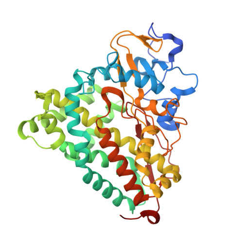
    Two key amino acids, Thr252 and Asp251, are known to be important for dioxygen activation by cytochrome P450cam. We have solved crystal structures of a critical intermediate, the ferrous dioxygen complex (Fe(II)-O2), of the wild-type P450cam and its mutants, D251N and T252A. The wild-type dioxygen complex structure is very much the same as reported previously (Schlichting, I., Berendzen, J., Chu, K., Stock, A. M., Maves, S. A., Benson, D. E., Sweet, R. M., Ringe, D., Petsko, G. A., and Sligar, S. G. (2000) Science 287, 1615-1622) with the exception of higher occupancy and a more ordered structure of the iron-linked dioxygen and two "catalytic" water molecules that form part of a proton relay system to the iron-linked dioxygen. Due to of the altered conformation of the I helix groove these two waters are missing in the D251N dioxygen complex which explains its lower catalytic activity and slower proton transfer to the dioxygen ligand. Similarly, the T252A mutation was expected to disrupt the active site solvent structure leading to hydrogen peroxide formation rather than substrate hydroxylation. Unexpectedly, however, the two "catalytic" waters are retained in the T252A mutant. Based on these findings, we propose that the Thr(252) accepts a hydrogen bond from the hydroperoxy (Fe(III)-OOH) intermediate that promotes the second protonation on the distal oxygen atom, leading to O-O bond cleavage and compound I formation.


  • Organizational Affiliation
    • Department of Molecular Biology & Biochemistry, University of California, Irvine 92697-3900, USA. snagano@riken.jp

Macromolecule Content 

  • Total Structure Weight: 95.21 kDa 
  • Atom Count: 7,055 
  • Modeled Residue Count: 812 
  • Deposited Residue Count: 830 
  • Unique protein chains: 1

Macromolecules

Find similar proteins by:|  3D Structure
Entity ID: 1
MoleculeChains  Sequence LengthOrganismDetailsImage
Cytochrome P450-cam
A, B
415Pseudomonas putidaMutation(s): 2 
Gene Names: CAMC
EC: 1.14.15.1
UniProt
Find proteins for P00183 (Pseudomonas putida)
Explore P00183 
Go to UniProtKB:  P00183
Entity Groups
Sequence Clusters30% Identity50% Identity70% Identity90% Identity95% Identity100% Identity
UniProt GroupP00183
Sequence Annotations
Expand
Reference Sequence

Small Molecules

Ligands 5 Unique
IDChains Name / Formula / InChI Key2D Diagram3D Interactions
HEM

Query on HEM


Download:Ideal Coordinates CCD File
E [auth A],
I [auth B]
PROTOPORPHYRIN IX CONTAINING FE
C34 H32 Fe N4 O4
KABFMIBPWCXCRK-RGGAHWMASA-L
CAM

Query on CAM


Download:Ideal Coordinates CCD File
G [auth A],
K [auth B]
CAMPHOR
C10 H16 O
DSSYKIVIOFKYAU-XCBNKYQSSA-N
TRS

Query on TRS


Download:Ideal Coordinates CCD File
L [auth B]2-AMINO-2-HYDROXYMETHYL-PROPANE-1,3-DIOL
C4 H12 N O3
LENZDBCJOHFCAS-UHFFFAOYSA-O
K

Query on K


Download:Ideal Coordinates CCD File
C [auth A],
D [auth A],
H [auth B]
POTASSIUM ION
K
NPYPAHLBTDXSSS-UHFFFAOYSA-N
OXY

Query on OXY


Download:Ideal Coordinates CCD File
F [auth A],
J [auth B]
OXYGEN MOLECULE
O2
MYMOFIZGZYHOMD-UHFFFAOYSA-N

Experimental Data & Validation

Experimental Data

  • Method: X-RAY DIFFRACTION
  • Resolution: 1.90 Å
  • R-Value Free:  0.235 (Depositor), 0.228 (DCC) 
  • R-Value Work:  0.191 (Depositor), 0.185 (DCC) 
  • R-Value Observed: 0.191 (Depositor) 
Space Group: P 1 21 1
Unit Cell:
Length ( Å )Angle ( ˚ )
a = 67.01α = 90
b = 62.13β = 90.4
c = 95.06γ = 90
Software Package:
Software NamePurpose
CNSrefinement
SCALEPACKdata scaling
BEASTphasing

Structure Validation

View Full Validation Report


Ligand Structure Quality Assessment 


Entry History 

Deposition Data

Revision History  (Full details and data files)

  • Version 1.0: 2005-07-05
    Type: Initial release
  • Version 1.1: 2008-04-30
    Changes: Version format compliance
  • Version 1.2: 2011-07-13
    Changes: Version format compliance
  • Version 1.3: 2017-10-11
    Changes: Refinement description
  • Version 1.4: 2021-11-10
    Changes: Database references, Derived calculations
  • Version 1.5: 2023-10-25
    Changes: Data collection, Refinement description