2A11 | pdb_00002a11

Crystal Structure of Nuclease Domain of Ribonuclase III from Mycobacterium Tuberculosis


Experimental Data Snapshot

  • Method: X-RAY DIFFRACTION
  • Resolution: 2.10 Å
  • R-Value Free: 
    0.286 (Depositor), 0.285 (DCC) 
  • R-Value Work: 
    0.259 (Depositor), 0.259 (DCC) 
  • R-Value Observed: 
    0.286 (Depositor) 

wwPDB Validation 3D Report Full Report

Validation slider image for 2A11

This is version 1.5 of the entry. See complete history

Literature

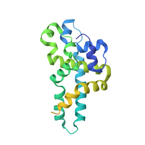
Structure of the nuclease domain of ribonuclease III from M. tuberculosis at 2.1 A

Akey, D.L.Berger, J.M.

(2005) Protein Sci 14: 2744-2750

  • DOI: https://doi.org/10.1110/ps.051665905
  • Primary Citation Related Structures: 
    2A11

  • PubMed Abstract: 

    RNase III enzymes are a highly conserved family of proteins that specifically cleave double-stranded (ds)RNA. These proteins are involved in a diverse group of functions, including ribosomal RNA processing, mRNA maturation and decay, snRNA and snoRNA processing, and RNA interference. Here we report the crystal structure of the nuclease domain of RNase III from the pathogen Mycobacterium tuberculosis. Although globally similar to other RNase III folds, this structure has some features not observed in previously reported models. These include the presence of an additional metal ion near the catalytic site, as well as conserved secondary structural elements that are proposed to have functional roles in the recognition of dsRNAs.


  • Organizational Affiliation
    • Department of Molecular and Cellular Biology, University of California, Berkeley, Berkeley, CA 94720, USA.

Macromolecule Content 

  • Total Structure Weight: 25.64 kDa 
  • Atom Count: 1,321 
  • Modeled Residue Count: 154 
  • Deposited Residue Count: 242 
  • Unique protein chains: 1

Macromolecules

Find similar proteins by:|  3D Structure
Entity ID: 1
MoleculeChains  Sequence LengthOrganismDetailsImage
Ribonuclease III242Mycobacterium tuberculosisMutation(s): 0 
Gene Names: rnc
EC: 3.1.26.3
UniProt
Find proteins for P9WH03 (Mycobacterium tuberculosis (strain ATCC 25618 / H37Rv))
Explore P9WH03 
Go to UniProtKB:  P9WH03
Entity Groups
Sequence Clusters30% Identity50% Identity70% Identity90% Identity95% Identity100% Identity
UniProt GroupP9WH03
Sequence Annotations
Expand
Reference Sequence

Experimental Data & Validation

Experimental Data

  • Method: X-RAY DIFFRACTION
  • Resolution: 2.10 Å
  • R-Value Free:  0.286 (Depositor), 0.285 (DCC) 
  • R-Value Work:  0.259 (Depositor), 0.259 (DCC) 
  • R-Value Observed: 0.286 (Depositor) 
Space Group: P 43 21 2
Unit Cell:
Length ( Å )Angle ( ˚ )
a = 72.589α = 90
b = 72.589β = 90
c = 96.013γ = 90
Software Package:
Software NamePurpose
Blu-Icedata collection
DENZOdata reduction
SCALEPACKdata scaling
SOLVEphasing
CNSrefinement

Structure Validation

View Full Validation Report


Entry History 

Deposition Data

Revision History  (Full details and data files)

  • Version 1.0: 2005-07-05
    Type: Initial release
  • Version 1.1: 2008-04-30
    Changes: Version format compliance
  • Version 1.2: 2011-07-13
    Changes: Derived calculations, Version format compliance
  • Version 1.3: 2011-11-16
    Changes: Atomic model
  • Version 1.4: 2017-10-11
    Changes: Refinement description
  • Version 1.5: 2024-02-14
    Changes: Data collection, Database references, Derived calculations