Download Mendeley
The Structure of Escherichia coli MscL and Its Dimer Formation in Nanodiscs.
Rasmussen, T., Bahner, J.I., Flegler, V.J., Hove, T.T., Kraft, C., Rasmussen, A., Bottcher, B.(2026) J Mol Biol 438: 169967-169967
- PubMed: 42551783 Search on PubMed
- DOI: https://doi.org/10.1016/j.jmb.2026.169967
- Primary Citation Related Structures:
28YC - PubMed Abstract:
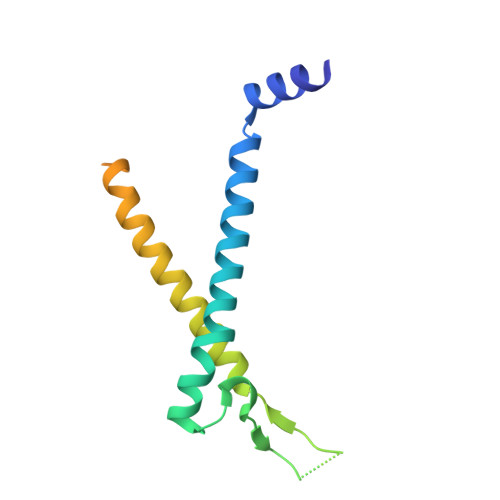
Mechanosensitive channels of large conductance (MscL) are essential bacterial safety valves that prevent osmotic lysis by releasing solutes in response to membrane tension. Despite extensive functional studies on Escherichia coli MscL (EcMscL), its high-resolution structure remained unknown. Using cryo-electron microscopy, we present an experimental structure of EcMscL reconstituted in nanodiscs at 3.1 Å resolution. The structure reveals a pentameric assembly with a narrow hydrophobic gate at the cytosolic side and a periplasmic cavity, consistent with the canonical MscL-fold. Differences to earlier published crystal structures of MscL from other organisms are in the less conserved periplasmic loop. We observe a previously unreported dimeric association of EcMscL pentamers, mediated by residues 61-63 in the periplasmic loop. This dimeric interface is located at the periplasmic side and provides a structural basis for the formation of higher-order clusters. The observed arrangement enables a fluid-like, mosaic packing of channels with centre-to-centre distances of 5.9-9 nm, consistent with biophysical and imaging data. These findings provide a structural framework for understanding cluster organisation of EcMscL that modulates its activity in cellular stress response.
- Julius-Maximilians-Universität Würzburg, Biocenter and Rudolf-Virchow-Center, Josef-Schneider-Str. 2, Gebäude D15, 97080 Würzburg, Germany.
Organizational Affiliation: