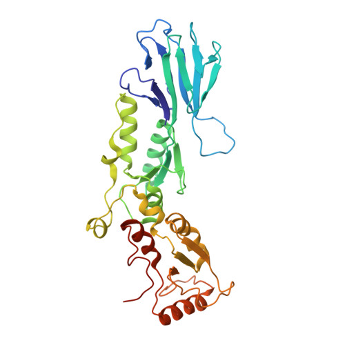
26PJ | pdb_000026pj

X-ray crystal structure of R28D mutant of the human NBS1 FHA-BRCT1-BRCT2 domain

  • Classification: CELL CYCLE
  • Organism(s): Homo sapiens
  • Expression System: Escherichia coli
  • Mutation(s): Yes 

  • Deposited: 2026-05-09 Released: 2026-10-28 
  • Deposition Author(s): Ite, K., Unno, M.
  • Funding Organization(s): Japan Agency for Medical Research and Development (AMED)

Experimental Data Snapshot

  • Method: X-RAY DIFFRACTION
  • Resolution: 2.84 Å
  • R-Value Free: 
    0.224 (Depositor), 0.221 (DCC) 
  • R-Value Work: 
    0.184 (Depositor), 0.189 (DCC) 

Starting Model: experimental
View more details

wwPDB Validation 3D Report Full Report

Validation slider image for 26PJ

This is version 1.0 of the entry. See complete history. 

Literature

X-ray structure of the human Nijmegen breakage syndrome forkhead-associated/BRCA1 C-terminal domain and computational docking of candidate radiosensitizers.

Unno, M., Kitanishi, K., Ite, K., Ishihara, M., Sugiyama, R., Komuro, S., Suzuki, K., Tauchi, H.

(2026) Acta Crystallogr F Struct Biol Commun 

  • DOI: https://doi.org/10.1107/S2053230X26010204
  • Primary Citation Related Structures: 
    26BE, 26PJ

  • PubMed Abstract: 

    Nijmegen breakage syndrome 1 (NBS1) forms the MRE11-RAD50-NBS1 complex and is essential for homologous recombination repair of DNA double-strand breaks. NBS1 regulates the localization and activity of the MRN complex. The NBS1 forkhead-associated (FHA) domain represents a potential target for the development of radiosensitizers. However, detailed structural information on the N-terminal region of human NBS1 (hNBS1) is still lacking. Here, the X-ray structure of the hNBS1 FHA-BRCA1 C-terminal (BRCT1)-BRCT2 domains is reported and ligand binding is evaluated using computational docking. A candidate radiosensitizer was predicted to bind at the tip of the FHA domain, indicating a potential interaction site. Substitution of Arg28 with aspartic acid within the FHA domain altered the orientation of neighboring side chains and modified the surface electrostatic properties, supporting its structural and functional importance. These findings provide a structural basis for targeting NBS1 and may facilitate the development of radiosensitizers.


  • Organizational Affiliation: 
    • Department of Applied Science and Engineering, Graduate School of Science and Engineering, Ibaraki University, Hitachi, Ibaraki 316-8511, Japan.

Macromolecule Content 

  • Total Structure Weight: 74.73 kDa 
  • Atom Count: 5,085 
  • Modeled Residue Count: 650 
  • Deposited Residue Count: 668 
  • Unique protein chains: 1

Macromolecules

Find similar proteins by:|  3D Structure
Entity ID: 1
MoleculeChains  Sequence LengthOrganismDetailsImage
Nibrin
A, B
334Homo sapiensMutation(s): 1 
Gene Names: NBN, NBS, NBS1, P95
UniProt & NIH Common Fund Data Resources
Find proteins for O60934 (Homo sapiens)
Explore O60934 
Go to UniProtKB:  O60934
PHAROS:  O60934
GTEx:  ENSG00000104320 
Entity Groups
Sequence Clusters30% Identity50% Identity70% Identity90% Identity95% Identity100% Identity
UniProt GroupO60934
Sequence Annotations
Expand
Reference Sequence

Experimental Data & Validation

Experimental Data

  • Method: X-RAY DIFFRACTION
  • Resolution: 2.84 Å
  • R-Value Free:  0.224 (Depositor), 0.221 (DCC) 
  • R-Value Work:  0.184 (Depositor), 0.189 (DCC) 
Space Group: C 2 2 21
Unit Cell:
Length ( Å )Angle ( ˚ )
a = 157.721α = 90
b = 182.384β = 90
c = 122.635γ = 90
Software Package:
Software NamePurpose
REFMACrefinement
XDSdata reduction
Aimlessdata scaling
MOLREPphasing

Structure Validation

View Full Validation Report


Entry History 

& Funding Information

Deposition Data


Funding OrganizationLocationGrant Number
Japan Agency for Medical Research and Development (AMED)Japan--

Revision History  (Full details and data files)

  • Version 1.0: 2026-10-28
    Type: Initial release