24IO | pdb_000024io

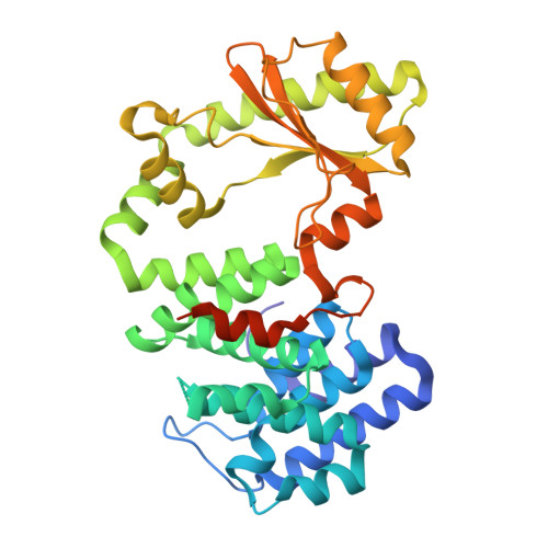
Crystal structure of the RelSeq N-terminal domain from Streptococcus equisimilis in complex with pppGpp


Experimental Data Snapshot

  • Method: X-RAY DIFFRACTION
  • Resolution: 3.20 Å
  • R-Value Free: 
    0.299 (Depositor), 0.294 (DCC) 
  • R-Value Work: 
    0.242 (Depositor), 0.239 (DCC) 
  • R-Value Observed: 
    0.245 (Depositor) 

Starting Model: experimental
View more details

wwPDB Validation 3D Report Full Report

Validation slider image for 24IO

Ligand Structure Quality Assessment 


This is version 1.1 of the entry. See complete history

Literature

Structural Basis of pppGpp Binding to the N-Terminal Domain of the Bifunctional RelA/SpoT Homolog Rel Seq : Crystal Structure and MD Analysis.

Korban, S.A.Spiridonova, Z.A.Kasatsky, P.S.Shvetsov, A.V.Gurzhiy, V.V.Paleskava, A.Kulminskaya, A.A.Konevega, A.L.Vinogradova, D.S.

(2026) Int J Mol Sci 27

  • DOI: https://doi.org/10.3390/ijms27125509
  • Primary Citation Related Structures: 
    24IO

  • PubMed Abstract: 

    RelA/SpoT homologue family enzymes participate in controlling the cellular levels of the alarmone (p)ppGpp, thereby activating the stringent response and promoting survival under stress conditions. These proteins contain an N-terminal catalytic domain and a C-terminal regulatory domain. They catalyze both the synthesis of ppGpp/pppGpp from ATP and GDP/GTP and their hydrolysis to GDP/GTP and pyrophosphate. Here, we report the crystal structure of the N-terminal domain of Rel from Streptococcus equisimilis in complex with pppGpp at 3.2 Å resolution. The asymmetric unit contains a dimer with asymmetric ligation: pppGpp occupies only the synthetase site in one monomer, whereas in the other monomer, it is bound in both the hydrolase and synthetase sites. The two monomers exhibit distinct conformational states, with pronounced rearrangements of the flexible loops surrounding the binding pockets, including the α2/α3 and α8/α9 loops that act as steric gates. Molecular dynamics simulations support the dual binding arrangement and reveal additional probable transient binding sites, including a region in the linker between hydrolase and synthetase subdomains. These findings provide a structural framework for understanding how pppGpp binding modulates the opposing catalytic activities of bifunctional Rel enzymes and suggest possible mechanisms for (p)ppGpp-mediated autoregulation.


  • Organizational Affiliation
    • Petersburg Nuclear Physics Institute Named by B.P. Konstantinov of National Research Centre "Kurchatov Institute", Gatchina 188300, Russia.

Macromolecule Content 

  • Total Structure Weight: 94.79 kDa 
  • Atom Count: 5,519 
  • Modeled Residue Count: 657 
  • Deposited Residue Count: 786 
  • Unique protein chains: 1

Macromolecules

Find similar proteins by:|  3D Structure
Entity ID: 1
MoleculeChains  Sequence LengthOrganismDetailsImage
Bifunctional (p)ppGpp synthase/hydrolase RelA
A, B
393Streptococcus dysgalactiae subsp. equisimilisMutation(s): 0 
Gene Names: relArel
EC: 2.7.6.5 (PDB Primary Data), 3.1.7.2 (PDB Primary Data)
UniProt
Find proteins for Q54089 (Streptococcus dysgalactiae subsp. equisimilis)
Explore Q54089 
Go to UniProtKB:  Q54089
Entity Groups
Sequence Clusters30% Identity50% Identity70% Identity90% Identity95% Identity100% Identity
UniProt GroupQ54089
Sequence Annotations
Expand
Reference Sequence

Small Molecules

Ligands 4 Unique
IDChains Name / Formula / InChI Key2D Diagram3D Interactions
0O2
(Subject of Investigation/LOI)

Query on 0O2


Download:Ideal Coordinates CCD File
P [auth A],
T [auth B],
U [auth B]
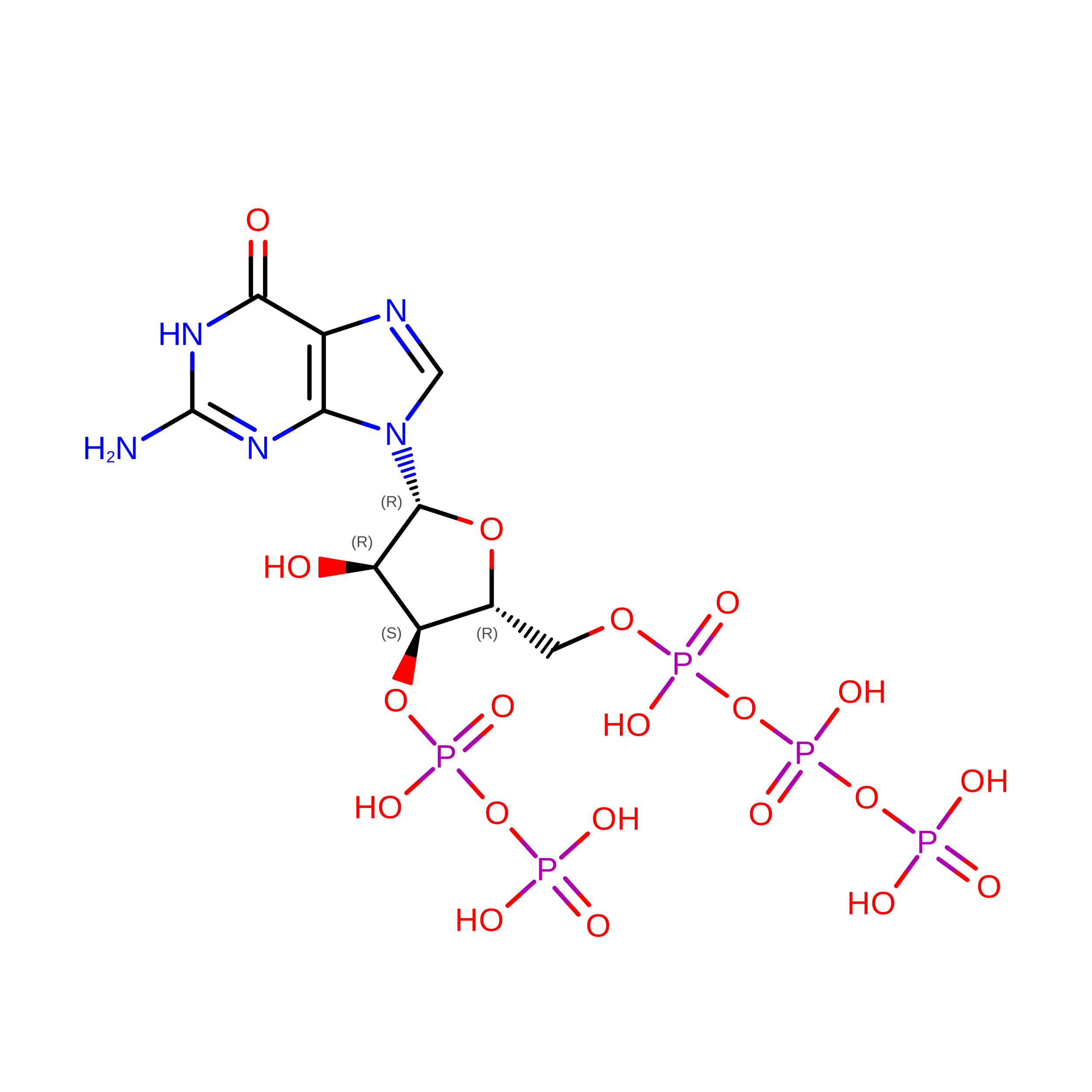
guanosine 5'-(tetrahydrogen triphosphate) 3'-(trihydrogen diphosphate)
C10 H18 N5 O20 P5
KCPMACXZAITQAX-UUOKFMHZSA-N
GOL

Query on GOL


Download:Ideal Coordinates CCD File
C [auth A]
D [auth A]
E [auth A]
F [auth A]
G [auth A]
C [auth A],
D [auth A],
E [auth A],
F [auth A],
G [auth A],
H [auth A],
I [auth A],
J [auth A],
K [auth A],
L [auth A],
M [auth A],
O [auth A],
Q [auth A],
R [auth A],
V [auth B]
GLYCEROL
C3 H8 O3
PEDCQBHIVMGVHV-UHFFFAOYSA-N
MN

Query on MN


Download:Ideal Coordinates CCD File
N [auth A],
S [auth B]
MANGANESE (II) ION
Mn
WAEMQWOKJMHJLA-UHFFFAOYSA-N
CL

Query on CL


Download:Ideal Coordinates CCD File
W [auth B]CHLORIDE ION
Cl
VEXZGXHMUGYJMC-UHFFFAOYSA-M

Experimental Data & Validation

Experimental Data

  • Method: X-RAY DIFFRACTION
  • Resolution: 3.20 Å
  • R-Value Free:  0.299 (Depositor), 0.294 (DCC) 
  • R-Value Work:  0.242 (Depositor), 0.239 (DCC) 
  • R-Value Observed: 0.245 (Depositor) 
Space Group: C 1 2 1
Unit Cell:
Length ( Å )Angle ( ˚ )
a = 173.751α = 90
b = 44.985β = 110.083
c = 126.433γ = 90
Software Package:
Software NamePurpose
PHENIXrefinement
CrysalisProdata collection
autoPROCdata processing
STARANISOdata scaling
PHENIXphasing
autoPROCdata reduction

Structure Validation

View Full Validation Report


Ligand Structure Quality Assessment 


Entry History 

& Funding Information

Deposition Data


Funding OrganizationLocationGrant Number
Russian Science FoundationRussian Federation23-74-10088
St. Petersburg State UniversityRussian Federation126022017738-9

Revision History  (Full details and data files)

  • Version 1.0: 2026-03-18
    Type: Initial release
  • Version 1.1: 2026-07-29
    Changes: Database references