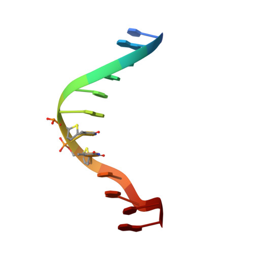
The crystal structure analysis of d(CGCGAASSCGCG)2, a synthetic DNA dodecamer duplex containing four 4'-thio-2'-deoxythymidine nucleotides.
Boggon, T.J., Hancox, E.L., McAuley-Hecht, K.E., Connolly, B.A., Hunter, W.N., Brown, T., Walker, R.T., Leonard, G.A.(1996) Nucleic Acids Res 24: 951-961
- PubMed: 8600465
- DOI: https://doi.org/10.1093/nar/24.5.951
- Primary Citation of Related Structures:
233D - PubMed Abstract:
The crystal structure refinement of the synthetic dodecamer d(CGCGAASSCGCG), where S = 4'-thio-2'-deoxythymidine, has converged at R=0.201 for 2605 reflections with F > 2sigma(F) in the resolution range 8.0-2.4 A for a model consisting of the dodecamer duplex and 66 water molecules. A comparison of its structure with that of the native dodecamer d(CGCGAATTCGCG) has revealed that the major differences between the two structures is a change in the conformation of the sugar-phosphate backbone in the regions at and adjacent to the positions of the modified nucleosides. Examination of the fine structural parameters for each of the structures reveals that the thiosugars adopt a C3'-exo conformation in d(CGCGAASSCGCG), rather than the approximate C1'-exo conformation found for the analogous sugars in the structure of d(CGCGAATTCGCG). The observed differences in structure between the two duplexes may help to explain the enhanced resistance to nuclease digestion of synthetic oligonucleotides containing 4'-thio-2'-deoxynucleotides.
Organizational Affiliation:
Department of Chemistry, University of Manchester, UK.


