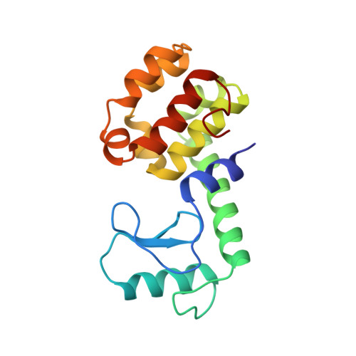
Context-dependent protein stabilization by methionine-to-leucine substitution shown in T4 lysozyme.
Lipscomb, L.A., Gassner, N.C., Snow, S.D., Eldridge, A.M., Baase, W.A., Drew, D.L., Matthews, B.W.(1998) Protein Sci 7: 765-773
- PubMed: 9541409 Search on PubMedSearch on PubMed Central
- DOI: https://doi.org/10.1002/pro.5560070326
- Primary Citation Related Structures:
230L, 231L, 232L, 233L, 234L - PubMed Abstract:
The substitution of methionines with leucines within the interior of a protein is expected to increase stability both because of a more favorable solvent transfer term as well as the reduced entropic cost of holding a leucine side chain in a defined position. Together, these two terms are expected to contribute about 1.4 kcal/mol to protein stability for each Met --> Leu substitution when fully buried. At the same time, this expected beneficial effect may be offset by steric factors due to differences in the shape of leucine and methionine. To investigate the interplay between these factors, all methionines in T4 lysozyme except at the amino-terminus were individually replaced with leucine. Of these mutants, M106L and M120L have stabilities 0.5 kcal/mol higher than wild-type T4 lysozyme, while M6L is significantly destabilized (-2.8 kcal/mol). M102L, described previously, is also destabilized (-0.9 kcal/mol). Based on this limited sample it appears that methionine-to-leucine substitutions can increase protein stability but only in a situation where the methionine side chain is fully or partially buried, yet allows the introduction of the leucine without concomitant steric interference. The variants, together with methionine-to-lysine substitutions at the same sites, follow the general pattern that substitutions at rigid, internal sites tend to be most destabilizing, whereas replacements at more solvent-exposed sites are better tolerated.
- Institute of Molecular Biology, Howard Hughes Medical Institute and Department of Chemistry, University of Oregon, Eugene 97403, USA.
Organizational Affiliation: