






 Download Mendeley
DPR epimerase complex, substrate-bound state
Wu, F., Gao, S., Rao, Z., Zhang, L.To be published.
Experimental Data Snapshot
wwPDB Validation 3D Report Full Report

Entity ID: 1 | |||||
|---|---|---|---|---|---|
| Molecule | Chains | Sequence Length | Organism | Details | Image |
| Decaprenylphosphoryl-beta-D-ribose oxidase | A [auth B], C [auth A] | 461 | Mycobacterium tuberculosis H37Rv | Mutation(s): 1 Gene Names: dprE1, Rv3790 EC: 1.1.98.3 | 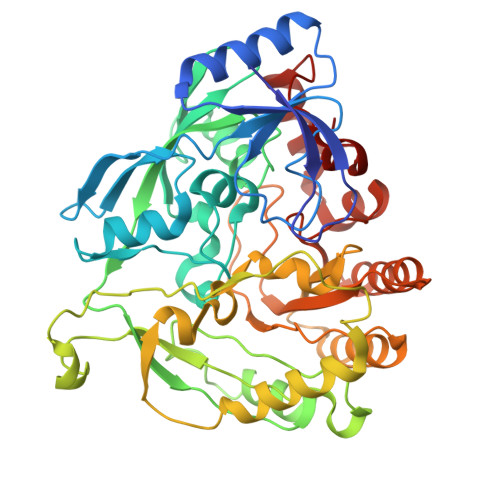 |
UniProt | |||||
Entity Groups | |||||
| Sequence Clusters | 30% Identity50% Identity70% Identity90% Identity95% Identity100% Identity | ||||
| UniProt Group | P9WJF1 | ||||
Sequence AnnotationsExpand | |||||
Reference Sequence | |||||
Entity ID: 2 | |||||
|---|---|---|---|---|---|
| Molecule | Chains | Sequence Length | Organism | Details | Image |
| Decaprenylphosphoryl-2-keto-beta-D-erythro-pentose reductase | B [auth D], D [auth C] | 254 | Mycobacterium tuberculosis H37Rv | Mutation(s): 0 Gene Names: dprE2, Rv3791, MTCY13D12.25 EC: 1.1.1.333 | 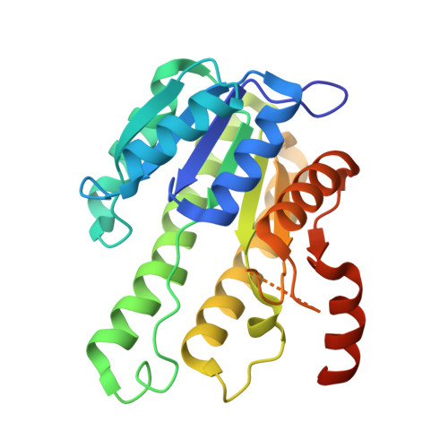 |
UniProt | |||||
Entity Groups | |||||
| Sequence Clusters | 30% Identity50% Identity70% Identity90% Identity95% Identity100% Identity | ||||
| UniProt Group | P9WGS9 | ||||
Sequence AnnotationsExpand | |||||
Reference Sequence | |||||
| Ligands 3 Unique | |||||
|---|---|---|---|---|---|
| ID | Chains | Name / Formula / InChI Key | 2D Diagram | 3D Interactions | |
| FAD (Subject of Investigation/LOI) Download:Ideal Coordinates CCD File | E [auth B], H [auth A] | FLAVIN-ADENINE DINUCLEOTIDE C27 H33 N9 O15 P2 VWWQXMAJTJZDQX-UYBVJOGSSA-N | 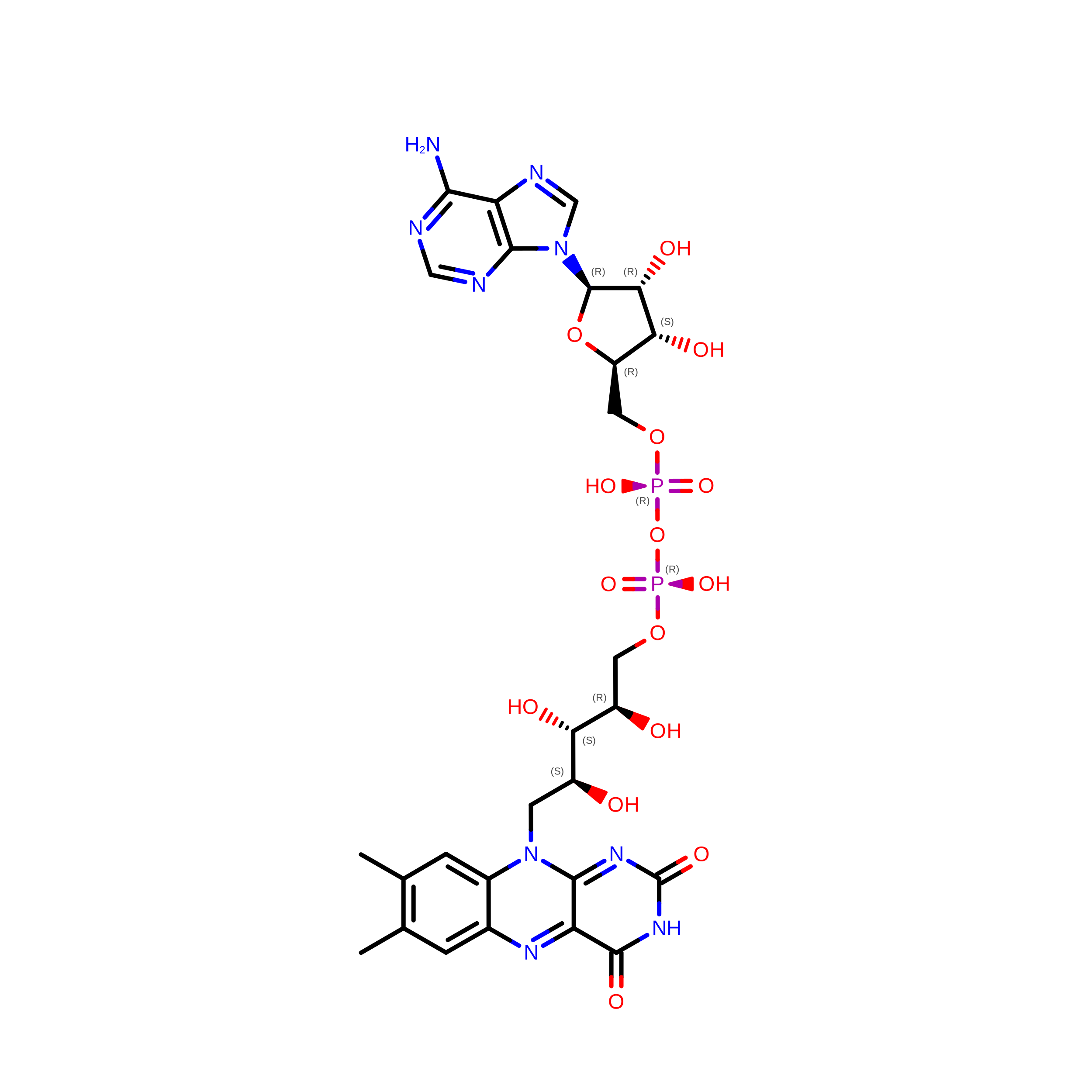 | ||
| NAI (Subject of Investigation/LOI) Download:Ideal Coordinates CCD File | G [auth D], J [auth C] | 1,4-DIHYDRONICOTINAMIDE ADENINE DINUCLEOTIDE C21 H29 N7 O14 P2 BOPGDPNILDQYTO-NNYOXOHSSA-N | 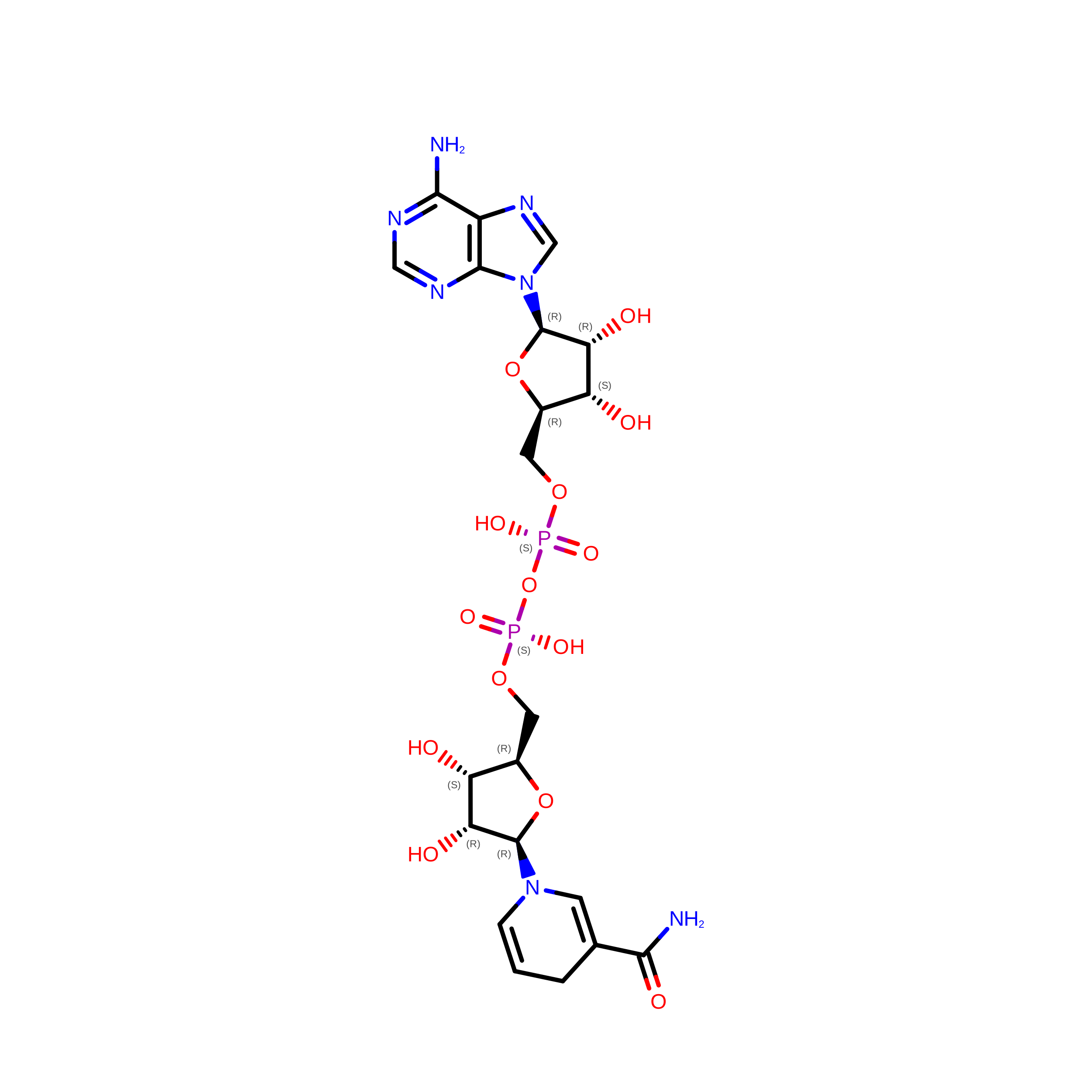 | ||
| A1L5J( Subject of Investigation/LOI) Download:Ideal Coordinates CCD File | F [auth B], I [auth A] | [(2~{E})-3,7-dimethylocta-2,6-dienyl] [(2~{S},3~{R},4~{S},5~{R})-5-(hydroxymethyl)-3,4-bis(oxidanyl)oxolan-2-yl] hydrogen phosphate C15 H27 O8 P YSLCRGJRVMNMDN-XDBUCDCOSA-N |  | ||
| Task | Software Package | Version |
|---|---|---|
| RECONSTRUCTION | cryoSPARC | |
| MODEL REFINEMENT | PHENIX |
| Funding Organization | Location | Grant Number |
|---|---|---|
| National Natural Science Foundation of China (NSFC) | China | 32394011 |








