21FG | pdb_000021fg

Cryo-EM structure of the human P2X3 receptor in the ATP- and sivopixant-bound closed state


Experimental Data Snapshot

  • Method: ELECTRON MICROSCOPY
  • Resolution: 3.34 Å
  • Aggregation State: PARTICLE 
  • Reconstruction Method: SINGLE PARTICLE 

wwPDB Validation 3D Report Full Report

Validation slider image for 21FG

This is version 1.1 of the entry. See complete history

Literature

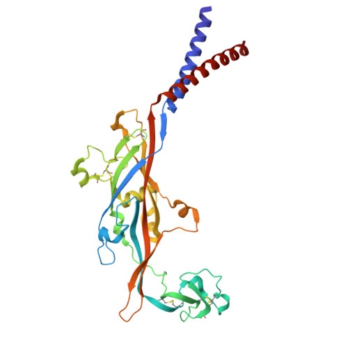
Structure of the human P2X3 receptor reveals the basis for subtype-selective inhibition by sivopixant.

Zhao, Z.Wang, D.P.Zhang, X.Gao, Y.Xu, H.Teng, X.Shen, C.Chen, J.Zhang, J.Guo, C.R.Hattori, M.

(2026) PLoS Biol 24: e3003777-e3003777

  • DOI: https://doi.org/10.1371/journal.pbio.3003777
  • Primary Citation Related Structures: 
    21DX, 21FG

  • PubMed Abstract: 

    P2X receptors are ATP-gated cation channels, and the P2X3 subtype plays crucial roles in peripheral sensory neurons, including in chronic pain and chronic cough. Accordingly, P2X3 receptors have attracted substantial interest as a therapeutic target. Gefapixant, a negative allosteric modulator (NAM) of P2X3 receptors, has been approved in some countries for the treatment of chronic cough; however, its limited selectivity for P2X3 homomers over P2X2/P2X3 heteromers is associated with taste disturbance as a prominent adverse effect. These limitations have motivated the development of next-generation NAMs with improved subtype selectivity, but their subtype-specific allosteric inhibition mechanisms are unclear. Here, we report the cryo-EM structure of the human P2X3 receptor in complex with ATP and the P2X3-selective next-generation NAM sivopixant, an investigational drug. Sivopixant binds to an allosteric site at the portal of the central pocket in the extracellular domain, and structure-based mutational analysis by electrophysiology identifies key residues required for sivopixant-dependent inhibition of human P2X3 receptors. Structural comparisons across P2X subtypes, together with patch-clamp analyses of gain-of-function mutants that confer sensitivity to two investigational drugs, sivopixant and camlipixant, provided a broadly applicable structural framework for subtype selectivity. Furthermore, structural comparisons with apo and ATP-bound open states of P2X3 receptors, together with molecular dynamics simulations, revealed that sivopixant expands the upper-body domain to suppress the lower-body movements required for channel activation, thereby preventing channel opening even in the presence of ATP.


  • Organizational Affiliation
    • State Key Laboratory of Genetics and Development of Complex Phenotypes, Collaborative Innovation Center of Genetics and Development, Department of Physiology and Neurobiology, School of Life Sciences, Fudan University, Shanghai, China.

Macromolecule Content 

  • Total Structure Weight: 114.97 kDa 
  • Atom Count: 7,521 
  • Modeled Residue Count: 981 
  • Deposited Residue Count: 981 
  • Unique protein chains: 1

Macromolecules

Find similar proteins by:|  3D Structure
Entity ID: 1
MoleculeChains  Sequence LengthOrganismDetailsImage
P2X purinoceptor 3
A, B, C
327Homo sapiensMutation(s): 0 
Gene Names: P2RX3
UniProt & NIH Common Fund Data Resources
Find proteins for P56373 (Homo sapiens)
Explore P56373 
Go to UniProtKB:  P56373
PHAROS:  P56373
GTEx:  ENSG00000109991 
Entity Groups
Sequence Clusters30% Identity50% Identity70% Identity90% Identity95% Identity100% Identity
UniProt GroupP56373
Glycosylation
Glycosylation Sites: 3Go to GlyGen: P56373-1
Sequence Annotations
Expand
Reference Sequence

Small Molecules

Ligands 3 Unique
IDChains Name / Formula / InChI Key2D Diagram3D Interactions
A1E2N
(Subject of Investigation/LOI)

Query on A1E2N


Download:Ideal Coordinates CCD File
D [auth A],
J [auth B],
O [auth C]
Sivopixant
C25 H22 Cl N5 O5
SKZQFKBIJUXXCG-INIZCTEOSA-N
ATP
(Subject of Investigation/LOI)

Query on ATP


Download:Ideal Coordinates CCD File
H [auth A],
I [auth A],
N [auth B]
ADENOSINE-5'-TRIPHOSPHATE
C10 H16 N5 O13 P3
ZKHQWZAMYRWXGA-KQYNXXCUSA-N
NAG

Query on NAG


Download:Ideal Coordinates CCD File
E [auth A]
F [auth A]
G [auth A]
K [auth B]
L [auth B]
E [auth A],
F [auth A],
G [auth A],
K [auth B],
L [auth B],
M [auth B],
P [auth C],
Q [auth C],
R [auth C]
2-acetamido-2-deoxy-beta-D-glucopyranose
C8 H15 N O6
OVRNDRQMDRJTHS-FMDGEEDCSA-N

Experimental Data & Validation

Experimental Data

  • Method: ELECTRON MICROSCOPY
  • Resolution: 3.34 Å
  • Aggregation State: PARTICLE 
  • Reconstruction Method: SINGLE PARTICLE 
EM Software:
TaskSoftware PackageVersion
RECONSTRUCTIONcryoSPARC

Structure Validation

View Full Validation Report


Entry History 

& Funding Information

Deposition Data


Funding OrganizationLocationGrant Number
National Natural Science Foundation of China (NSFC)China--

Revision History  (Full details and data files)

  • Version 1.0: 2026-04-01
    Type: Initial release
  • Version 1.1: 2026-05-06
    Changes: Data collection, Database references