21AK | pdb_000021ak

Cryo-EM structure of the E. coli ArnA hexamer


Experimental Data Snapshot

  • Method: ELECTRON MICROSCOPY
  • Resolution: 3.23 Å
  • Aggregation State: PARTICLE 
  • Reconstruction Method: SINGLE PARTICLE 

Starting Models: experimental
View more details

wwPDB Validation 3D Report Full Report

Validation slider image for 21AK

This is version 1.2 of the entry. See complete history

Literature

Cryo-EM reveals ArnA contamination during purification of a ciliary protein complex.

Jiang, X.Kikkawa, M.

(2026) Acta Crystallogr D Struct Biol 82: 404-410

  • DOI: https://doi.org/10.1107/S2059798326002846
  • Primary Citation Related Structures: 
    21AK

  • PubMed Abstract: 

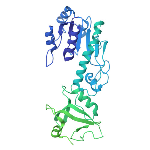
    Endogenous Escherichia coli proteins can co-purify with recombinant targets and dominate cryo-EM datasets, yet often escape detection during standard biochemical quality control. In parallel to the recent report by Caliseki and coworkers [Caliseki et al. (2025), Acta Cryst. D81, 545-557], we independently identified ArnA contamination while purifying a soluble, low-yield KIF17-IFT70 complex, ultimately obtaining a 3.23 Å resolution cryo-EM structure of ArnA rather than the intended target. Our results reinforce that ArnA enrichment reflects general features of His-tag affinity purification and can become particularly problematic in cryo-EM workflows when the intended target is low in yield or conformationally heterogeneous. By comparing biochemical behavior and cryo-EM outcomes, we outline why ArnA may evade SDS-PAGE and size-exclusion chromatography, and be underappreciated in routine mass spectrometry-based quality control, yet becomes structurally dominant in cryo-EM. These findings broaden the scope of the original study and highlight the need for early cryo-EM screening and improved contaminant awareness in structural biology.


  • Organizational Affiliation
    • Department of Cell Biology and Anatomy, Graduate School of Medicine, The University of Tokyo, Tokyo 113-0033, Japan.

Macromolecule Content 

  • Total Structure Weight: 892.63 kDa 
  • Atom Count: 28,592 
  • Modeled Residue Count: 3,612 
  • Deposited Residue Count: 7,920 
  • Unique protein chains: 1

Macromolecules

Find similar proteins by:|  3D Structure
Entity ID: 1
MoleculeChains  Sequence LengthOrganismDetailsImage
Bifunctional polymyxin resistance protein ArnA
A, B, C, D, E
A, B, C, D, E, F, G, H, I, J, K, L
660Escherichia coliMutation(s): 0 
Gene Names: arnApmrIyfbGb2255JW2249
EC: 2.1.2.13 (PDB Primary Data), 1.1.1.305 (PDB Primary Data)
UniProt
Find proteins for P77398 (Escherichia coli (strain K12))
Explore P77398 
Go to UniProtKB:  P77398
Entity Groups
Sequence Clusters30% Identity50% Identity70% Identity90% Identity95% Identity100% Identity
UniProt GroupP77398
Sequence Annotations
Expand
Reference Sequence

Experimental Data & Validation

Experimental Data

  • Method: ELECTRON MICROSCOPY
  • Resolution: 3.23 Å
  • Aggregation State: PARTICLE 
  • Reconstruction Method: SINGLE PARTICLE 
EM Software:
TaskSoftware PackageVersion
RECONSTRUCTIONcryoSPARCv4.7.1
MODEL REFINEMENTPHENIX
MODEL REFINEMENTCoot

Structure Validation

View Full Validation Report


Entry History 

& Funding Information

Deposition Data


Funding OrganizationLocationGrant Number
Japan Science and TechnologyJapanJPMJER2202
Japan Society for the Promotion of Science (JSPS)JapanJP21H05247
Japan Society for the Promotion of Science (JSPS)JapanJP24KF0141
Japan Society for the Promotion of Science (JSPS)JapanJP24K18106

Revision History  (Full details and data files)

  • Version 1.0: 2026-03-25
    Type: Initial release
  • Version 1.1: 2026-04-22
    Changes: Data collection, Database references
  • Version 1.2: 2026-05-13
    Changes: Data collection, Database references