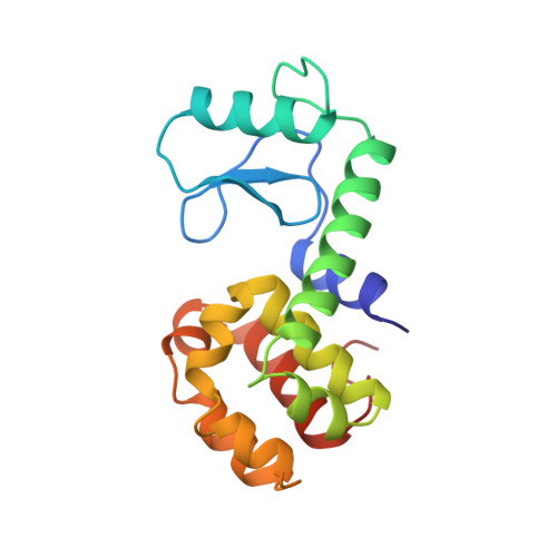
Protein structural plasticity exemplified by insertion and deletion mutants in T4 lysozyme.
Vetter, I.R., Baase, W.A., Heinz, D.W., Xiong, J.P., Snow, S., Matthews, B.W.(1996) Protein Sci 5: 2399-2415
- PubMed: 8976549
- DOI: https://doi.org/10.1002/pro.5560051203
- Primary Citation of Related Structures:
209L, 210L, 211L, 212L, 213L, 214L, 215L, 218L, 219L - PubMed Abstract:
To further investigate the ways in which proteins respond to changes in the length of the polypeptide chain, a series of 32 insertions and five deletions were made within nine different alpha-helices of T4 lysozyme. In most cases, the inserted amino acid was a single alanine, although in some instances up to four residues, not necessarily alanine, were used. Different insertions destabilized the protein by different amounts, ranging from approximately 1 to 6 kcal/mol. In one case, no protein could be obtained. An "extension" mutant in which the carboxy terminus of the molecule was extended by four alanines increased stability by 0.3 kcal/mol. For the deletions, the loss in stability ranged from approximately 3 to 5 kcal/mol. The structures of six insertion mutants, as well as one deletion mutant and the extension mutant, were determined, three in crystal forms nonisomorphous with wild type. In all cases, including previously described insertion mutants within a single alpha-helix, there appears to be a strong tendency to preserve the helix by translocating residues so that the effects of the insertion are propagated into a bend or loop at one end or the other of the helix. In three mutants, even the hydrophobic core was disrupted so as to permit the preservation of the alpha-helix containing the insertion. Translocation (or "register shift") was also observed for the deletion mutant, in this case a loop at the end of the helix being shortened. In general, when translocation occurs, the reduction in stability is only moderate, averaging 2.5 kcal/mol. Only in the most extreme cases does "bulging" or "looping-out" occur within the body of an alpha-helix, in which case the destabilization is substantial, averaging 4.9 kcal/mol. Looping-out can occur for insertions close to the end of a helix, in which case the destabilization is less severe, averaging 2.6 kcal/mol. Mutant A73-[AAA] as well as mutants R119-[A] and V131-[A], include shifts in the backbone of 3-6 A, extending over 20 residues or more. As a result, residues 114-142, which form a "cap" on the carboxy-terminal domain, undergo substantial reorganizations such that the interface between this "cap" and the rest of the protein is altered substantially. In the case of mutant A73-[AAA], two nearby alpha-helices, which form a bend of approximately 105 degrees in the wild-type structure, reorganize in the mutant structure to form a single, essentially straight helix. These structural responses to mutation demonstrate the plasticity of protein structures and illustrate ways in which their three-dimensional structures might changes during evolution.
Organizational Affiliation:
Institute of Molecular Biology, Howard Hughes Medical Institute, University of Oregon, Eugene 97403, USA.


