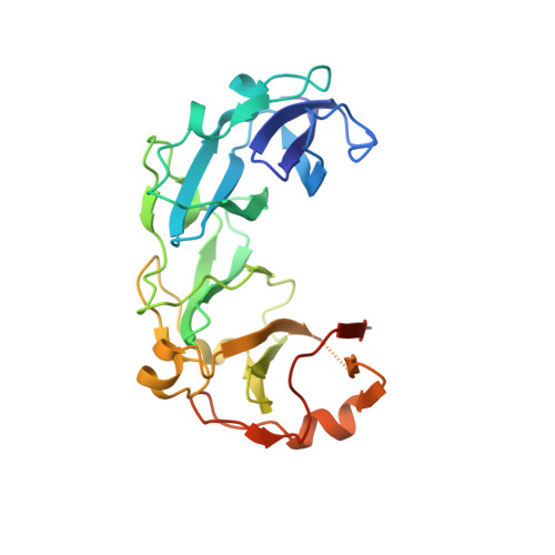
The Structural Basis for Substrate Specificity in DNA Topoisomerase IV.
Corbett, K.D., Schoeffler, A.J., Thomsen, N.D., Berger, J.M.(2005) J Mol Biology 351: 545-561
- PubMed: 16023670
- DOI: https://doi.org/10.1016/j.jmb.2005.06.029
- Primary Citation of Related Structures:
1ZVT, 1ZVU - PubMed Abstract:
Most bacteria possess two type IIA topoisomerases, DNA gyrase and topo IV, that together help manage chromosome integrity and topology. Gyrase primarily introduces negative supercoils into DNA, an activity mediated by the C-terminal domain of its DNA binding subunit (GyrA). Although closely related to gyrase, topo IV preferentially decatenates DNA and relaxes positive supercoils. Here we report the structure of the full-length Escherichia coli ParC dimer at 3.0 A resolution. The N-terminal DNA binding region of ParC is highly similar to that of GyrA, but the ParC dimer adopts a markedly different conformation. The C-terminal domain (CTD) of ParC is revealed to be a degenerate form of the homologous GyrA CTD, and is anchored to the top of the N-terminal domains in a configuration different from that thought to occur in gyrase. Biochemical assays show that the ParC CTD controls the substrate specificity of topo IV, likely by capturing DNA segments of certain crossover geometries. This work delineates strong mechanistic parallels between topo IV and gyrase, while explaining how structural differences between the two enzyme families have led to distinct activity profiles. These findings in turn explain how the structures and functions of bacterial type IIA topoisomerases have evolved to meet specific needs of different bacterial families for the control of chromosome superstructure.
- Department of Molecular and Cell Biology, 237 Hildebrand Hall #3206, University of California, Berkeley, Berkeley, CA 94720-3206, USA.
Organizational Affiliation:


