1YNQ | pdb_00001ynq

aldo-keto reductase AKR11C1 from Bacillus halodurans (holo form)


Experimental Data Snapshot

  • Method: X-RAY DIFFRACTION
  • Resolution: 1.30 Å
  • R-Value Free: 
    0.185 (Depositor), 0.185 (DCC) 
  • R-Value Work: 
    0.152 (Depositor), 0.151 (DCC) 
  • R-Value Observed: 
    0.153 (Depositor) 

wwPDB Validation 3D Report Full Report

Validation slider image for 1YNQ

Ligand Structure Quality Assessment 


This is version 2.1 of the entry. See complete history

Literature

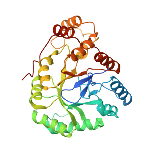
High-resolution Crystal Structure of AKR11C1 from Bacillus halodurans: An NADPH-dependent 4-Hydroxy-2,3-trans-nonenal Reductase

Marquardt, T.Kostrewa, D.Balakrishnan, R.Gasperina, A.Kambach, C.Podjarny, A.Winkler, F.K.Balendiran, G.K.Li, X.D.

(2005) J Mol Biology 354: 304-316

  • DOI: https://doi.org/10.1016/j.jmb.2005.09.067
  • Primary Citation Related Structures: 
    1YNP, 1YNQ

  • PubMed Abstract: 

    Aldo-keto reductase AKR11C1 from Bacillus halodurans, a new member of aldo-keto reductase (AKR) family 11, has been characterized structurally and biochemically. The structures of the apo and NADPH bound form of AKR11C1 have been solved to 1.25 A and 1.3 A resolution, respectively. AKR11C1 possesses a novel non-aromatic stacking interaction of an arginine residue with the cofactor, which may favor release of the oxidized cofactor. Our biochemical studies have revealed an NADPH-dependent activity of AKR11C1 with 4-hydroxy-2,3-trans-nonenal (HNE). HNE is a cytotoxic lipid peroxidation product, and detoxification in alkaliphilic bacteria, such as B.halodurans, plays a crucial role in survival. AKR11C1 could thus be part of the detoxification system, which ensures the well being of the microorganism. The very poor activity of AKR11C1 on standard, small substrates such as benzaldehyde or DL-glyeraldehyde is consistent with the observed, very open active site lacking a binding pocket for these substrates. In contrast, modeling of HNE with its aldehyde function suitably positioned in the active site suggests that its elongated hydrophobic tail occupies a groove defined by hydrophobic side-chains. Multiple sequence alignment of AKR11C1 with the highly homologous iolS and YqkF proteins shows a high level of conservation in this putative substrate-binding site. We suggest that AKR11C1 is the first structurally characterized member of a new class of AKRs with specificity for substrates with long aliphatic tails.


  • Organizational Affiliation
    • Biomolecular Research, Paul Scherrer Institut, 5232 Villigen, Switzerland.

Macromolecule Content 

  • Total Structure Weight: 74.65 kDa 
  • Atom Count: 5,564 
  • Modeled Residue Count: 586 
  • Deposited Residue Count: 634 
  • Unique protein chains: 1

Macromolecules

Find similar proteins by:|  3D Structure
Entity ID: 1
MoleculeChains  Sequence LengthOrganismDetailsImage
oxidoreductase
A, B
317Halalkalibacterium halodurans C-125Mutation(s): 0 
Gene Names: BH1011
UniProt
Find proteins for Q9KE47 (Halalkalibacterium halodurans (strain ATCC BAA-125 / DSM 18197 / FERM 7344 / JCM 9153 / C-125))
Explore Q9KE47 
Go to UniProtKB:  Q9KE47
Entity Groups
Sequence Clusters30% Identity50% Identity70% Identity90% Identity95% Identity100% Identity
UniProt GroupQ9KE47
Sequence Annotations
Expand
Reference Sequence

Oligosaccharides

Help  
Entity ID: 2
MoleculeChains Length2D Diagram GlycosylationD Interactions
beta-D-fructofuranose-(2-1)-alpha-D-glucopyranose
C
2N/A
Glycosylation Resources
GlyTouCan: G05551OP
GlyCosmos: G05551OP

Small Molecules

Ligands 4 Unique
IDChains Name / Formula / InChI Key2D Diagram3D Interactions
NDP

Query on NDP


Download:Ideal Coordinates CCD File
P [auth B]NADPH DIHYDRO-NICOTINAMIDE-ADENINE-DINUCLEOTIDE PHOSPHATE
C21 H30 N7 O17 P3
ACFIXJIJDZMPPO-NNYOXOHSSA-N
SO4

Query on SO4


Download:Ideal Coordinates CCD File
E [auth A]
F [auth A]
G [auth A]
H [auth A]
I [auth A]
E [auth A],
F [auth A],
G [auth A],
H [auth A],
I [auth A],
J [auth A],
K [auth A],
L [auth A],
M [auth B],
N [auth B],
O [auth B]
SULFATE ION
O4 S
QAOWNCQODCNURD-UHFFFAOYSA-L
GOL

Query on GOL


Download:Ideal Coordinates CCD File
Q [auth B],
R [auth B],
S [auth B]
GLYCEROL
C3 H8 O3
PEDCQBHIVMGVHV-UHFFFAOYSA-N
NA

Query on NA


Download:Ideal Coordinates CCD File
D [auth A]SODIUM ION
Na
FKNQFGJONOIPTF-UHFFFAOYSA-N

Biologically Interesting Molecules (External Reference) 

1 Unique

Experimental Data & Validation

Experimental Data

  • Method: X-RAY DIFFRACTION
  • Resolution: 1.30 Å
  • R-Value Free:  0.185 (Depositor), 0.185 (DCC) 
  • R-Value Work:  0.152 (Depositor), 0.151 (DCC) 
  • R-Value Observed: 0.153 (Depositor) 
Space Group: P 21 21 21
Unit Cell:
Length ( Å )Angle ( ˚ )
a = 75.122α = 90
b = 87.543β = 90
c = 105.434γ = 90
Software Package:
Software NamePurpose
REFMACrefinement
PDB_EXTRACTdata extraction
MAR345data collection
XDSdata scaling

Structure Validation

View Full Validation Report


Ligand Structure Quality Assessment 


Entry History 

Deposition Data

Revision History  (Full details and data files)

  • Version 1.0: 2005-12-06
    Type: Initial release
  • Version 1.1: 2008-04-01
    Changes: Version format compliance
  • Version 1.2: 2011-07-13
    Changes: Source and taxonomy, Version format compliance
  • Version 1.3: 2017-10-11
    Changes: Refinement description
  • Version 2.0: 2020-07-29
    Type: Remediation
    Reason: Carbohydrate remediation
    Changes: Atomic model, Data collection, Database references, Derived calculations, Non-polymer description, Structure summary
  • Version 2.1: 2024-03-13
    Changes: Data collection, Database references, Structure summary