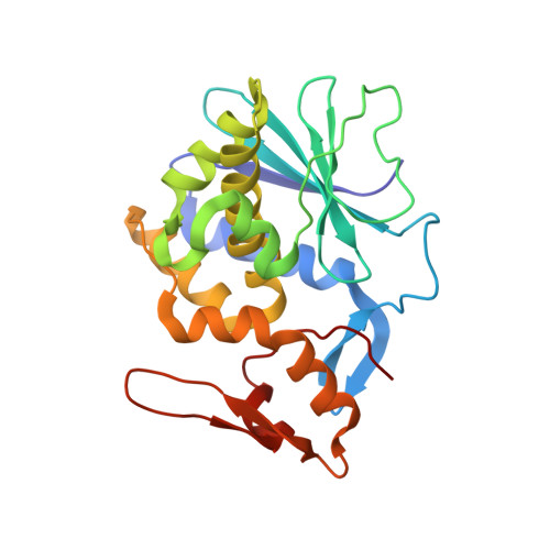
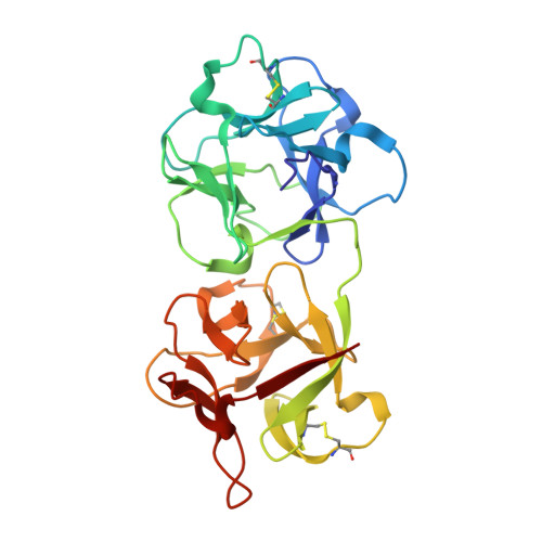
1YF8

Crystal structure of Himalayan mistletoe RIP reveals the presence of a natural inhibitor and a new functionally active sugar-binding site


Experimental Data Snapshot

  • Method: X-RAY DIFFRACTION
  • Resolution: 2.80 Å
  • R-Value Free: 0.287 
  • R-Value Work: 0.234 
  • R-Value Observed: 0.237 

Starting Model: experimental
View more details

wwPDB Validation   3D Report Full Report


This is version 2.1 of the entry. See complete history


Literature

Crystal structure of himalayan mistletoe ribosome-inactivating protein reveals the presence of a natural inhibitor and a new functionally active sugar-binding site.

Mishra, V.Bilgrami, S.Sharma, R.S.Kaur, P.Yadav, S.Krauspenhaar, R.Betzel, C.Voelter, W.Babu, C.R.Singh, T.P.

(2005) J Biol Chem 280: 20712-20721

  • DOI: https://doi.org/10.1074/jbc.M500735200
  • Primary Citation of Related Structures:  
    1YF8

  • PubMed Abstract: 

    Ribosome-inactivating proteins (RIPs) are toxins involved in plant defense. How the plant prevents autotoxicity is not yet fully understood. The present study is the first structural evidence of a naturally inhibited form of RIP from a plant. Himalayan mistletoe RIP (HmRIP) was purified from Viscum album leaves and crystallized with lactose. The structure was determined by the molecular replacement method and refined at 2.8-A resolution. The crystal structure revealed the presence of high quality non-protein electron density at the active site, into which a pteridine derivative (2-amino 4-isopropyl 6-carboxyl pteridine) was modeled. The carboxyl group of the ligand binds strongly with the key active site residue Arg(162), nullifies the positive charge required for catalysis, and thereby acts as a natural inhibitor. Lectin subunits of RIPs have two active sugar-binding sites present in 1alpha- and 2gamma-subdomains. A third functionally active site has been identified in the 1beta-subdomain of HmRIP. The 1beta-site is active despite the absence of conserved polar sugar-binding residues. Loss of these residues is compensated by the following: (i) the presence of an extended site where the penultimate sugar also interacts with the protein; (ii) the interactions of galactose with the protein main chain carbonyl and amide nitrogen atoms; (iii) the presence of a well defined pocket encircled by four walls; and (iv) a favorable stacking of the galactose ring with Tyr(66) besides the conserved Phe(75). The mode of sugar binding is also distinct at the 1alpha and 2gamma sugar-binding sites.


  • Organizational Affiliation

    Department of Biophysics, All India Institute of Medical Sciences, New Delhi 110 029, India.


Macromolecules
Find similar proteins by:  (by identity cutoff)  |  3D Structure
Entity ID: 1
MoleculeChains Sequence LengthOrganismDetailsImage
Beta-galactoside-specific lectin 4240Viscum albumMutation(s): 0 
EC: 3.2.2.22
UniProt
Find proteins for Q6ITZ3 (Viscum album)
Explore Q6ITZ3 
Go to UniProtKB:  Q6ITZ3
Entity Groups  
Sequence Clusters30% Identity50% Identity70% Identity90% Identity95% Identity100% Identity
UniProt GroupQ6ITZ3
Glycosylation
Glycosylation Sites: 1
Sequence Annotations
Expand
  • Reference Sequence
Find similar proteins by:  (by identity cutoff)  |  3D Structure
Entity ID: 2
MoleculeChains Sequence LengthOrganismDetailsImage
Beta-galactoside-specific lectin 4255Viscum albumMutation(s): 0 
EC: 3.2.2.22
UniProt
Find proteins for Q6ITZ3 (Viscum album)
Explore Q6ITZ3 
Go to UniProtKB:  Q6ITZ3
Entity Groups  
Sequence Clusters30% Identity50% Identity70% Identity90% Identity95% Identity100% Identity
UniProt GroupQ6ITZ3
Glycosylation
Glycosylation Sites: 3
Sequence Annotations
Expand
  • Reference Sequence
Oligosaccharides

Help

Entity ID: 3
MoleculeChains Length2D Diagram Glycosylation3D Interactions
2-acetamido-2-deoxy-beta-D-glucopyranose-(1-4)-2-acetamido-2-deoxy-beta-D-glucopyranose
C
2N-Glycosylation
Glycosylation Resources
GlyTouCan:  G42666HT
GlyCosmos:  G42666HT
GlyGen:  G42666HT
Entity ID: 4
MoleculeChains Length2D Diagram Glycosylation3D Interactions
beta-D-mannopyranose-(1-3)-alpha-D-mannopyranose-(1-4)-2-acetamido-2-deoxy-beta-D-glucopyranose-(1-4)-2-acetamido-2-deoxy-beta-D-glucopyranose
D
4N-Glycosylation
Glycosylation Resources
GlyTouCan:  G24199JF
GlyCosmos:  G24199JF
GlyGen:  G24199JF
Entity ID: 5
MoleculeChains Length2D Diagram Glycosylation3D Interactions
alpha-D-mannopyranose-(1-4)-2-acetamido-2-deoxy-beta-D-glucopyranose-(1-4)-2-acetamido-2-deoxy-beta-D-glucopyranose
E
3N-Glycosylation
Glycosylation Resources
GlyTouCan:  G62182OO
GlyCosmos:  G62182OO
GlyGen:  G62182OO
Entity ID: 6
MoleculeChains Length2D Diagram Glycosylation3D Interactions
beta-D-galactopyranose-(1-4)-beta-D-glucopyranose
F, G
2N/A
Glycosylation Resources
GlyTouCan:  G84224TW
GlyCosmos:  G84224TW
GlyGen:  G84224TW
Small Molecules
Ligands 3 Unique
IDChains Name / Formula / InChI Key2D Diagram3D Interactions
P6C
Query on P6C

Download Ideal Coordinates CCD File 
I [auth A]2-AMINO-4-ISOPROPYL-PTERIDINE-6-CARBOXYLIC ACID
C10 H11 N5 O2
GPHPGFBRJYCUDO-UHFFFAOYSA-N
NAG
Query on NAG

Download Ideal Coordinates CCD File 
H [auth A]2-acetamido-2-deoxy-beta-D-glucopyranose
C8 H15 N O6
OVRNDRQMDRJTHS-FMDGEEDCSA-N
GAL
Query on GAL

Download Ideal Coordinates CCD File 
J [auth B]beta-D-galactopyranose
C6 H12 O6
WQZGKKKJIJFFOK-FPRJBGLDSA-N
Biologically Interesting Molecules (External Reference) 1 Unique
Experimental Data & Validation

Experimental Data

  • Method: X-RAY DIFFRACTION
  • Resolution: 2.80 Å
  • R-Value Free: 0.287 
  • R-Value Work: 0.234 
  • R-Value Observed: 0.237 
  • Space Group: P 65 2 2
Unit Cell:
Length ( Å )Angle ( ˚ )
a = 109.422α = 90
b = 109.422β = 90
c = 309.798γ = 120
Software Package:
Software NamePurpose
REFMACrefinement
DENZOdata reduction
SCALEPACKdata scaling
AMoREphasing

Structure Validation

View Full Validation Report


Entry History 

Deposition Data

Revision History  (Full details and data files)

  • Version 1.0: 2005-03-08
    Type: Initial release
  • Version 1.1: 2008-04-30
    Changes: Version format compliance
  • Version 1.2: 2011-07-13
    Changes: Non-polymer description, Version format compliance
  • Version 2.0: 2020-07-29
    Type: Remediation
    Reason: Carbohydrate remediation
    Changes: Advisory, Atomic model, Data collection, Derived calculations, Structure summary
  • Version 2.1: 2023-10-25
    Changes: Data collection, Database references, Refinement description, Structure summary