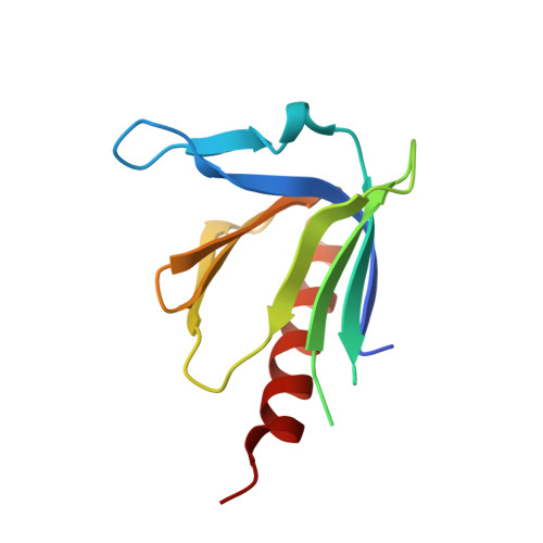
1XOD

Crystal structure of X. tropicalis Spred1 EVH-1 domain


Experimental Data Snapshot

  • Method: X-RAY DIFFRACTION
  • Resolution: 1.15 Å
  • R-Value Free: 0.177 
  • R-Value Work: 0.154 
  • R-Value Observed: 0.155 

Starting Models: experimental
View more details

wwPDB Validation   3D Report Full Report


This is version 1.3 of the entry. See complete history


Literature

1.15A Crystal structure of the X. tropicalis Spred1 EVH1 domain suggests a fourth distinct peptide-binding mechanism within the EVH1 family

Harmer, N.J.Sivak, J.M.Amaya, E.Blundell, T.L.

(2005) FEBS Lett 579: 1161-1166

  • DOI: https://doi.org/10.1016/j.febslet.2004.11.114
  • Primary Citation of Related Structures:  
    1XOD

  • PubMed Abstract: 

    The recently described Spred protein family has been implicated in the modulation of receptor tyrosine kinase signalling. We report the crystal structure of the Enabled/vasodilator-stimulated phosphoprotein homology-1 (EVH1) domain from Xenopus tropicalis Spred1, solved to 1.15 A resolution. This structure confirms that the Spred EVH1 adopts the pleckstrin-homology fold, with a similar secondary structure to Enabled. A translation of one of the peptide-binding groove beta-strands narrows this groove, whilst one end of the groove shows structural flexibility. We propose that Spred1 will bind peptides that are less proline-rich than other EVH1 domains, with conformational changes indicating an induced fit.


  • Organizational Affiliation

    Department of Biochemistry, 80 Tennis Court Road, Cambridge, CB2 1GA, UK. nic@cryst.bioc.cam.ac.uk


Macromolecules
Find similar proteins by:  (by identity cutoff)  |  3D Structure
Entity ID: 1
MoleculeChains Sequence LengthOrganismDetailsImage
Spred1
A, B
118Xenopus tropicalisMutation(s): 0 
Gene Names: Spred1
UniProt
Find proteins for Q66JG9 (Xenopus tropicalis)
Explore Q66JG9 
Go to UniProtKB:  Q66JG9
Entity Groups  
Sequence Clusters30% Identity50% Identity70% Identity90% Identity95% Identity100% Identity
UniProt GroupQ66JG9
Sequence Annotations
Expand
  • Reference Sequence
Small Molecules
Ligands 1 Unique
IDChains Name / Formula / InChI Key2D Diagram3D Interactions
GOL
Query on GOL

Download Ideal Coordinates CCD File 
C [auth B]GLYCEROL
C3 H8 O3
PEDCQBHIVMGVHV-UHFFFAOYSA-N
Experimental Data & Validation

Experimental Data

  • Method: X-RAY DIFFRACTION
  • Resolution: 1.15 Å
  • R-Value Free: 0.177 
  • R-Value Work: 0.154 
  • R-Value Observed: 0.155 
  • Space Group: P 1 21 1
Unit Cell:
Length ( Å )Angle ( ˚ )
a = 32.811α = 90
b = 38.193β = 95.68
c = 79.907γ = 90
Software Package:
Software NamePurpose
REFMACrefinement
HKL-2000data reduction
SCALEPACKdata scaling
AMoREphasing

Structure Validation

View Full Validation Report


Entry History 

Deposition Data

Revision History  (Full details and data files)

  • Version 1.0: 2005-01-25
    Type: Initial release
  • Version 1.1: 2008-04-30
    Changes: Version format compliance
  • Version 1.2: 2011-07-13
    Changes: Non-polymer description, Version format compliance
  • Version 1.3: 2023-08-23
    Changes: Data collection, Database references, Derived calculations, Refinement description