1WCQ | pdb_00001wcq

Mutagenesis of the Nucleophilic Tyrosine in a Bacterial Sialidase to Phenylalanine.


Experimental Data Snapshot

  • Method: X-RAY DIFFRACTION
  • Resolution: 2.10 Å
  • R-Value Free: 
    0.238 (Depositor), 0.238 (DCC) 
  • R-Value Work: 
    0.175 (Depositor), 0.177 (DCC) 
  • R-Value Observed: 
    0.178 (Depositor) 

Starting Model: experimental
View more details

wwPDB Validation 3D Report Full Report

Validation slider image for 1WCQ

Ligand Structure Quality Assessment 


This is version 1.4 of the entry. See complete history

Literature

Two Nucleophilic Mutants of the Micromonospora Viridifaciens Sialidase Operate with Retention of Configuration by Two Different Mechanisms.

Watson, J.N.Newstead, S.Narine, A.A.Taylor, G.Bennet, A.J.

(2005) Chembiochem 6: 1439

  • DOI: https://doi.org/10.1002/cbic.200500114
  • Primary Citation Related Structures: 
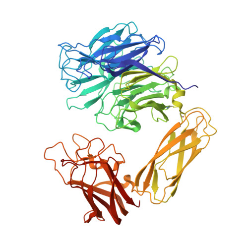
    1WCQ

  • PubMed Abstract: 

    Mutants of the Micromonospora viridifaciens sialidase, Y370E and Y370F, are catalytically active retaining enzymes that operate by different mechanisms. Previous substitutions with smaller amino acids, including Y370D, yielded inverting sialidases. At least one water molecule can fit into the active-site cavity of this mutant and act as a nucleophile from the face opposite the leaving group (Biochemistry 2003, 42, 12 682). Thus, addition of a CH(2) unit (Asp versus Glu) changes the mechanism from inversion back to retention of configuration. Based on Brønsted beta(lg) values, it is proposed that the Y370E mutant reacts by a double-displacement mechanism (beta(lg) on k(cat)/K(m) -0.36+/-0.04) with Glu370 acting as the nucleophile. However, the Y370F mutant (beta(lg) on k(cat)/K(m) -0.79+/-0.12) reacts via a dissociative transition state. The crystal structure of the Y370F mutant complexed with 2-deoxy-2,3-dehydro-N-acetylneuraminic acid shows no significant active-site perturbation relative to the wild-type enzyme.


  • Organizational Affiliation
    • Department of Chemistry, Simon Fraser University, University Drive, Burnaby, BC, Canada.

Macromolecule Content 

  • Total Structure Weight: 194.44 kDa 
  • Atom Count: 14,833 
  • Modeled Residue Count: 1,801 
  • Deposited Residue Count: 1,803 
  • Unique protein chains: 1

Macromolecules

Find similar proteins by:|  3D Structure
Entity ID: 1
MoleculeChains  Sequence LengthOrganismDetailsImage
SIALIDASE
A, B, C
601Micromonospora viridifaciensMutation(s): 1 
EC: 3.2.1.18
UniProt
Find proteins for Q02834 (Micromonospora viridifaciens)
Explore Q02834 
Go to UniProtKB:  Q02834
Entity Groups
Sequence Clusters30% Identity50% Identity70% Identity90% Identity95% Identity100% Identity
UniProt GroupQ02834
Sequence Annotations
Expand
Reference Sequence

Small Molecules

Ligands 3 Unique
IDChains Name / Formula / InChI Key2D Diagram3D Interactions
DAN

Query on DAN


Download:Ideal Coordinates CCD File
G [auth A],
L [auth B],
P [auth C]
2-DEOXY-2,3-DEHYDRO-N-ACETYL-NEURAMINIC ACID
C11 H17 N O8
JINJZWSZQKHCIP-UFGQHTETSA-N
GOL

Query on GOL


Download:Ideal Coordinates CCD File
E [auth A]
F [auth A]
I [auth B]
J [auth B]
K [auth B]
E [auth A],
F [auth A],
I [auth B],
J [auth B],
K [auth B],
N [auth C],
O [auth C]
GLYCEROL
C3 H8 O3
PEDCQBHIVMGVHV-UHFFFAOYSA-N
NA

Query on NA


Download:Ideal Coordinates CCD File
D [auth A],
H [auth B],
M [auth C]
SODIUM ION
Na
FKNQFGJONOIPTF-UHFFFAOYSA-N

Experimental Data & Validation

Experimental Data

  • Method: X-RAY DIFFRACTION
  • Resolution: 2.10 Å
  • R-Value Free:  0.238 (Depositor), 0.238 (DCC) 
  • R-Value Work:  0.175 (Depositor), 0.177 (DCC) 
  • R-Value Observed: 0.178 (Depositor) 
Space Group: P 32 2 1
Unit Cell:
Length ( Å )Angle ( ˚ )
a = 143.258α = 90
b = 143.258β = 90
c = 160.25γ = 120
Software Package:
Software NamePurpose
REFMACrefinement
MOSFLMdata reduction
SCALAdata scaling
AMoREphasing

Structure Validation

View Full Validation Report


Ligand Structure Quality Assessment 


Entry History 

Deposition Data

Revision History  (Full details and data files)

  • Version 1.0: 2005-10-12
    Type: Initial release
  • Version 1.1: 2011-07-13
    Changes: Advisory, Version format compliance
  • Version 1.2: 2020-07-29
    Type: Remediation
    Reason: Carbohydrate remediation
    Changes: Data collection, Derived calculations, Other
  • Version 1.3: 2023-12-13
    Changes: Data collection, Database references, Refinement description, Structure summary
  • Version 1.4: 2024-10-23
    Changes: Structure summary