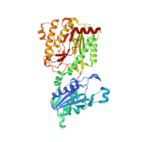
1W7V

ZnMg substituted aminopeptidase P from E. coli


Experimental Data Snapshot

  • Method: X-RAY DIFFRACTION
  • Resolution: 2.00 Å
  • R-Value Free: 0.180 
  • R-Value Work: 0.156 
  • R-Value Observed: 0.157 

wwPDB Validation   3D Report Full Report


This is version 1.5 of the entry. See complete history


Literature

Structural and Functional Implications of Metal Ion Selection in Aminopeptidase P, a Metalloprotease with a Dinuclear Metal Center.

Graham, S.C.Bond, C.S.Freeman, H.C.Guss, J.M.

(2005) Biochemistry 44: 13820

  • DOI: https://doi.org/10.1021/bi0512849
  • Primary Citation of Related Structures:  
    1W2M, 1W7V, 1WBQ, 1WL6, 1WL9, 1WLR, 2BH3, 2BHA, 2BHB, 2BHC, 2BHD, 2BN7

  • PubMed Abstract: 

    The effect of metal substitution on the activity and structure of the aminopeptidase P (APPro) from Escherichia coli has been investigated. Measurements of activity in the presence of Mn2+, Mg2+, Zn2+, Na+, and Ca2+ show that significant activity is seen only in the Mn-bound form of the enzyme. The addition of Zn2+ to [MnMn(APPro)] is strongly inhibitory. Crystal structures of [MnMn(APPro)], [MgMg(APPro)], [ZnZn(APPro)], [ZnMg(APPro)], [Ca_(APPro)], [Na_(APPro)], and [apo(APPro)] were determined. The structures of [Ca_(APPro)] and [Na_(APPro)] have a single metal atom at their active site. Surprisingly, when a tripeptide substrate (ValProLeu) was soaked into [Na_(APPro)] crystals in the presence of 200 mM Mg2+, the structure had substrate, but no metal, bound at the active site. The structure of apo APPro complexed with ValProLeu shows that the N-terminal amino group of a substrate can be bound at the active site by carboxylate side chains that normally bind the second metal atom, providing a model for substrate binding in a single-metal active enzyme. Structures of [MnMn(APPro)] and [ZnZn(APPro)] complexes of ProLeu, a product inhibitor, in the presence of excess Zn reveal a third metal-binding site, formed by two conserved His residues and the dipeptide inhibitor. A Zn atom bound at such a site would stabilize product binding and enhance inhibition.


  • Organizational Affiliation

    School of Molecular and Microbial Biosciences, University of Sydney, NSW 2006, Australia.


Macromolecules
Find similar proteins by:  (by identity cutoff)  |  3D Structure
Entity ID: 1
MoleculeChains Sequence LengthOrganismDetailsImage
XAA-PRO AMINOPEPTIDASE
A, B, C, D
440Escherichia coliMutation(s): 0 
EC: 3.4.11.9
UniProt
Find proteins for P15034 (Escherichia coli (strain K12))
Explore P15034 
Go to UniProtKB:  P15034
Entity Groups  
Sequence Clusters30% Identity50% Identity70% Identity90% Identity95% Identity100% Identity
UniProt GroupP15034
Sequence Annotations
Expand
  • Reference Sequence

Find similar proteins by:  Sequence   |   3D Structure  

Entity ID: 2
MoleculeChains Sequence LengthOrganismDetailsImage
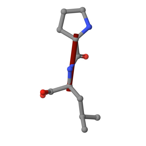
PEPTIDE VAL-PRO-LEU
E, F, G, H
3synthetic constructMutation(s): 0 
Sequence Annotations
Expand
  • Reference Sequence
Small Molecules
Ligands 3 Unique
IDChains Name / Formula / InChI Key2D Diagram3D Interactions
ZN
Query on ZN

Download Ideal Coordinates CCD File 
I [auth A],
L [auth B],
P [auth C],
S [auth D]
ZINC ION
Zn
PTFCDOFLOPIGGS-UHFFFAOYSA-N
CL
Query on CL

Download Ideal Coordinates CCD File 
K [auth A],
O [auth B],
R [auth C],
V [auth D]
CHLORIDE ION
Cl
VEXZGXHMUGYJMC-UHFFFAOYSA-M
MG
Query on MG

Download Ideal Coordinates CCD File 
J [auth A]
M [auth B]
N [auth B]
Q [auth C]
T [auth D]
J [auth A],
M [auth B],
N [auth B],
Q [auth C],
T [auth D],
U [auth D]
MAGNESIUM ION
Mg
JLVVSXFLKOJNIY-UHFFFAOYSA-N
Modified Residues  1 Unique
IDChains TypeFormula2D DiagramParent
OCS
Query on OCS
A, B, C, D
L-PEPTIDE LINKINGC3 H7 N O5 SCYS
Experimental Data & Validation

Experimental Data

  • Method: X-RAY DIFFRACTION
  • Resolution: 2.00 Å
  • R-Value Free: 0.180 
  • R-Value Work: 0.156 
  • R-Value Observed: 0.157 
  • Space Group: C 1 2 1
Unit Cell:
Length ( Å )Angle ( ˚ )
a = 111.731α = 90
b = 236.527β = 106.34
c = 137.997γ = 90
Software Package:
Software NamePurpose
REFMACrefinement
DENZOdata reduction
SCALEPACKdata scaling
AMoREphasing

Structure Validation

View Full Validation Report


Entry History 

Deposition Data

Revision History  (Full details and data files)

  • Version 1.0: 2005-09-29
    Type: Initial release
  • Version 1.1: 2011-07-13
    Changes: Advisory, Refinement description, Version format compliance
  • Version 1.2: 2019-05-08
    Changes: Data collection, Derived calculations, Experimental preparation, Other
  • Version 1.3: 2019-10-09
    Changes: Data collection, Other
  • Version 1.4: 2020-07-29
    Changes: Derived calculations, Source and taxonomy
  • Version 1.5: 2023-12-13
    Changes: Data collection, Database references, Refinement description