1W7V | pdb_00001w7v

ZnMg substituted aminopeptidase P from E. coli


Experimental Data Snapshot

  • Method: X-RAY DIFFRACTION
  • Resolution: 2.00 Å
  • R-Value Free: 
    0.180 (Depositor), 0.192 (DCC) 
  • R-Value Work: 
    0.156 (Depositor), 0.169 (DCC) 
  • R-Value Observed: 
    0.157 (Depositor) 

Starting Model: experimental
View more details

wwPDB Validation 3D Report Full Report

Validation slider image for 1W7V

This is version 1.5 of the entry. See complete history

Literature

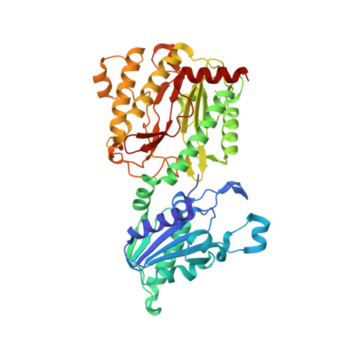
Structural and Functional Implications of Metal Ion Selection in Aminopeptidase P, a Metalloprotease with a Dinuclear Metal Center.

Graham, S.C.Bond, C.S.Freeman, H.C.Guss, J.M.

(2005) Biochemistry 44: 13820

  • DOI: https://doi.org/10.1021/bi0512849
  • Primary Citation Related Structures: 
    1W2M, 1W7V, 1WBQ, 1WL6, 1WL9, 1WLR, 2BH3, 2BHA, 2BHB, 2BHC, 2BHD, 2BN7

  • PubMed Abstract: 

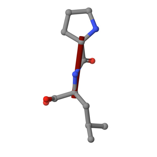
    The effect of metal substitution on the activity and structure of the aminopeptidase P (APPro) from Escherichia coli has been investigated. Measurements of activity in the presence of Mn2+, Mg2+, Zn2+, Na+, and Ca2+ show that significant activity is seen only in the Mn-bound form of the enzyme. The addition of Zn2+ to [MnMn(APPro)] is strongly inhibitory. Crystal structures of [MnMn(APPro)], [MgMg(APPro)], [ZnZn(APPro)], [ZnMg(APPro)], [Ca_(APPro)], [Na_(APPro)], and [apo(APPro)] were determined. The structures of [Ca_(APPro)] and [Na_(APPro)] have a single metal atom at their active site. Surprisingly, when a tripeptide substrate (ValProLeu) was soaked into [Na_(APPro)] crystals in the presence of 200 mM Mg2+, the structure had substrate, but no metal, bound at the active site. The structure of apo APPro complexed with ValProLeu shows that the N-terminal amino group of a substrate can be bound at the active site by carboxylate side chains that normally bind the second metal atom, providing a model for substrate binding in a single-metal active enzyme. Structures of [MnMn(APPro)] and [ZnZn(APPro)] complexes of ProLeu, a product inhibitor, in the presence of excess Zn reveal a third metal-binding site, formed by two conserved His residues and the dipeptide inhibitor. A Zn atom bound at such a site would stabilize product binding and enhance inhibition.


  • Organizational Affiliation
    • School of Molecular and Microbial Biosciences, University of Sydney, NSW 2006, Australia.

Macromolecule Content 

  • Total Structure Weight: 201.03 kDa 
  • Atom Count: 15,192 
  • Modeled Residue Count: 1,765 
  • Deposited Residue Count: 1,772 
  • Unique protein chains: 2

Macromolecules

Find similar proteins by:|  3D Structure
Entity ID: 1
MoleculeChains  Sequence LengthOrganismDetailsImage
XAA-PRO AMINOPEPTIDASE
A, B, C, D
440Escherichia coliMutation(s): 0 
EC: 3.4.11.9
UniProt
Find proteins for P15034 (Escherichia coli (strain K12))
Explore P15034 
Go to UniProtKB:  P15034
Entity Groups
Sequence Clusters30% Identity50% Identity70% Identity90% Identity95% Identity100% Identity
UniProt GroupP15034
Sequence Annotations
Expand
Reference Sequence
Find similar proteins by:  Sequence   |   3D Structure  
Entity ID: 2
MoleculeChains  Sequence LengthOrganismDetailsImage
PEPTIDE VAL-PRO-LEU
E, F, G, H
3synthetic constructMutation(s): 0 

Small Molecules

Ligands 3 Unique
IDChains Name / Formula / InChI Key2D Diagram3D Interactions
ZN

Query on ZN


Download:Ideal Coordinates CCD File
I [auth A],
L [auth B],
P [auth C],
S [auth D]
ZINC ION
Zn
PTFCDOFLOPIGGS-UHFFFAOYSA-N
CL

Query on CL


Download:Ideal Coordinates CCD File
K [auth A],
O [auth B],
R [auth C],
V [auth D]
CHLORIDE ION
Cl
VEXZGXHMUGYJMC-UHFFFAOYSA-M
MG

Query on MG


Download:Ideal Coordinates CCD File
J [auth A]
M [auth B]
N [auth B]
Q [auth C]
T [auth D]
J [auth A],
M [auth B],
N [auth B],
Q [auth C],
T [auth D],
U [auth D]
MAGNESIUM ION
Mg
JLVVSXFLKOJNIY-UHFFFAOYSA-N
Modified Residues  1 Unique
IDChains TypeFormula2D DiagramParent
OCS
Query on OCS
A, B, C, D
L-PEPTIDE LINKINGC3 H7 N O5 SCYS

Experimental Data & Validation

Experimental Data

  • Method: X-RAY DIFFRACTION
  • Resolution: 2.00 Å
  • R-Value Free:  0.180 (Depositor), 0.192 (DCC) 
  • R-Value Work:  0.156 (Depositor), 0.169 (DCC) 
  • R-Value Observed: 0.157 (Depositor) 
Space Group: C 1 2 1
Unit Cell:
Length ( Å )Angle ( ˚ )
a = 111.731α = 90
b = 236.527β = 106.34
c = 137.997γ = 90
Software Package:
Software NamePurpose
REFMACrefinement
DENZOdata reduction
SCALEPACKdata scaling
AMoREphasing

Structure Validation

View Full Validation Report


Entry History 

Deposition Data

Revision History  (Full details and data files)

  • Version 1.0: 2005-09-29
    Type: Initial release
  • Version 1.1: 2011-07-13
    Changes: Advisory, Refinement description, Version format compliance
  • Version 1.2: 2019-05-08
    Changes: Data collection, Derived calculations, Experimental preparation, Other
  • Version 1.3: 2019-10-09
    Changes: Data collection, Other
  • Version 1.4: 2020-07-29
    Changes: Derived calculations, Source and taxonomy
  • Version 1.5: 2023-12-13
    Changes: Data collection, Database references, Refinement description