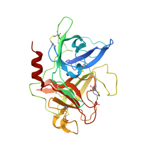
1W12

UROKINASE TYPE PLASMINOGEN ACTIVATOR


Experimental Data Snapshot

  • Method: X-RAY DIFFRACTION
  • Resolution: 2.40 Å
  • R-Value Free: 0.261 
  • R-Value Work: 0.199 
  • R-Value Observed: 0.199 

wwPDB Validation   3D Report Full Report


This is version 1.3 of the entry. See complete history


Literature

Crystals of Urokinase Type Plasminogen Activator Complexes Reveal the Binding Mode of Peptidomimetic Inhibitors.

Zeslawska, E.Jacob, U.Schweinitz, A.Coombs, G.Bode, W.Madison, E.

(2003) J Mol Biol 328: 109

  • DOI: https://doi.org/10.1016/s0022-2836(03)00267-5
  • Primary Citation of Related Structures:  
    1W0Z, 1W10, 1W11, 1W12, 1W13, 1W14

  • PubMed Abstract: 

    Urokinase type plasminogen activator (uPA), a trypsin-like serine proteinase, plays an important role in normal tissue re-modelling, cell adhesion, and cell motility. In addition, studies utilizing normal animals and potent, selective uPA inhibitors or genetically modified mice that lack functional uPA genes have demonstrated that uPA can significantly enhance tumor initiation, growth, progression and metastasis, strongly suggesting that this enzyme may be a promising anti-cancer target. We have investigated the structure-activity relationship (SAR) of peptidomimetic inhibitors of uPA and solved high resolution X-ray structures of key, lead small molecule inhibitors (e.g. phenethylsulfonamidino(P4)-D-seryl(P3)-L-alanyl(P2)-L-argininal(P1) and derivatives thereof) in complex with the uPA proteinase domain. These potent inhibitors are highly selective for uPA. The non-natural D-seryl residue present at the P3 position in these inhibitors contributes substantially to both potency and selectivity because, due to its D-configuration, its side-chain binds in the S4 pocket to interact with the uPA unique residues Leu97b and His99. Additional potency and selectivity can be achieved by optimizing the inhibitor P4 residue to bind a pocket, known as S1sub or S1beta, that is adjacent to the primary specificity pocket of uPA.


  • Organizational Affiliation

    Pedagogical University, Department of Chemistry, Podchorazych 2, 30-084 Cracow, Poland.


Macromolecules
Find similar proteins by:  (by identity cutoff)  |  3D Structure
Entity ID: 1
MoleculeChains Sequence LengthOrganismDetailsImage
UROKINASE-TYPE PLASMINOGEN ACTIVATORA [auth U]247Homo sapiensMutation(s): 0 
EC: 3.4.21.73
UniProt & NIH Common Fund Data Resources
Find proteins for P00749 (Homo sapiens)
Explore P00749 
Go to UniProtKB:  P00749
PHAROS:  P00749
GTEx:  ENSG00000122861 
Entity Groups  
Sequence Clusters30% Identity50% Identity70% Identity90% Identity95% Identity100% Identity
UniProt GroupP00749
Sequence Annotations
Expand
  • Reference Sequence
Small Molecules
Ligands 1 Unique
IDChains Name / Formula / InChI Key2D Diagram3D Interactions
SL1
Query on SL1

Download Ideal Coordinates CCD File 
B [auth U]N-((1S)-4-{[AMINO(IMINO)METHYL]AMINO}-1-FORMYLBUTYL)-2-{(3R)-3-[(BENZYLSULFONYL)AMINO]-2-OXO-5-PHENYL-2,3-DIHYDRO-1H-1,4-BENZODIAZEPIN-1-YL}ACETAMIDE
C30 H33 N7 O5 S
JVZSQVNCJHGRDE-NEKDWFFYSA-N
Binding Affinity Annotations 
IDSourceBinding Affinity
SL1 PDBBind:  1W12 Ki: 14 (nM) from 1 assay(s)
Experimental Data & Validation

Experimental Data

  • Method: X-RAY DIFFRACTION
  • Resolution: 2.40 Å
  • R-Value Free: 0.261 
  • R-Value Work: 0.199 
  • R-Value Observed: 0.199 
  • Space Group: P 41 21 2
Unit Cell:
Length ( Å )Angle ( ˚ )
a = 55.006α = 90
b = 55.006β = 90
c = 151.609γ = 90
Software Package:
Software NamePurpose
CNSrefinement

Structure Validation

View Full Validation Report


Entry History 

Deposition Data

  • Released Date: 2008-05-20 
  • Deposition Author(s): Jacob, U.

Revision History  (Full details and data files)

  • Version 1.0: 2008-05-20
    Type: Initial release
  • Version 1.1: 2011-05-08
    Changes: Version format compliance
  • Version 1.2: 2011-07-13
    Changes: Version format compliance
  • Version 1.3: 2019-05-22
    Changes: Data collection, Derived calculations, Other, Refinement description