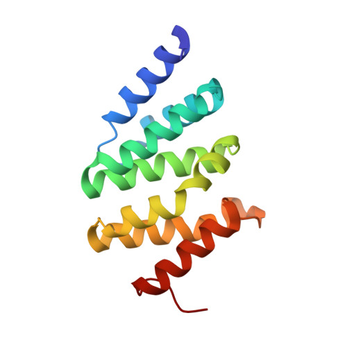
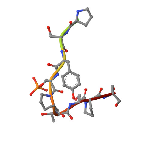
1SZA

The RNA polymerase II CTD in mRNA processing: beta-turn recognition and beta-spiral model


Experimental Data Snapshot

  • Method: X-RAY DIFFRACTION
  • Resolution: 2.20 Å
  • R-Value Free: 0.257 
  • R-Value Work: 0.211 
  • R-Value Observed: 0.211 

wwPDB Validation   3D Report Full Report


This is version 1.4 of the entry. See complete history


Literature

Recognition of RNA polymerase II carboxy-terminal domain by 3'-RNA-processing factors.

Meinhart, A.Cramer, P.

(2004) Nature 430: 223-226

  • DOI: https://doi.org/10.1038/nature02679
  • Primary Citation of Related Structures:  
    1SZ9, 1SZA

  • PubMed Abstract: 

    During transcription, RNA polymerase (Pol) II synthesizes eukaryotic messenger RNA. Transcription is coupled to RNA processing by the carboxy-terminal domain (CTD) of Pol II, which consists of up to 52 repeats of the sequence Tyr 1-Ser 2-Pro 3-Thr 4-Ser 5-Pro 6-Ser 7 (refs 1, 2). After phosphorylation, the CTD binds tightly to a conserved CTD-interacting domain (CID) present in the proteins Pcf11 and Nrd1, which are essential and evolutionarily conserved factors for polyadenylation-dependent and -independent 3'-RNA processing, respectively. Here we describe the structure of a Ser 2-phosphorylated CTD peptide bound to the CID domain of Pcf11. The CTD motif Ser 2-Pro 3-Thr 4-Ser 5 forms a beta-turn that binds to a conserved groove in the CID domain. The Ser 2 phosphate group does not make direct contact with the CID domain, but may be recognized indirectly because it stabilizes the beta-turn with an additional hydrogen bond. Iteration of the peptide structure results in a compact beta-spiral model of the CTD. The model suggests that, during the mRNA transcription-processing cycle, compact spiral regions in the CTD are unravelled and regenerated in a phosphorylation-dependent manner.


  • Organizational Affiliation

    Department of Chemistry and Biochemistry, Gene Center, University of Munich, 81377 Munich, Germany.


Macromolecules
Find similar proteins by:  (by identity cutoff)  |  3D Structure
Entity ID: 1
MoleculeChains Sequence LengthOrganismDetailsImage
PCF11 protein
A, B, C
144Saccharomyces cerevisiaeMutation(s): 0 
Gene Names: PCF11YDR228CYD9934.13C
UniProt
Find proteins for P39081 (Saccharomyces cerevisiae (strain ATCC 204508 / S288c))
Explore P39081 
Go to UniProtKB:  P39081
Entity Groups  
Sequence Clusters30% Identity50% Identity70% Identity90% Identity95% Identity100% Identity
UniProt GroupP39081
Sequence Annotations
Expand
  • Reference Sequence

Find similar proteins by:  Sequence   |   3D Structure  

Entity ID: 2
MoleculeChains Sequence LengthOrganismDetailsImage
CTD-peptideD [auth Z]14N/AMutation(s): 0 
Entity Groups  
Sequence Clusters30% Identity50% Identity70% Identity90% Identity95% Identity100% Identity
Sequence Annotations
Expand
  • Reference Sequence
Small Molecules
Modified Residues  1 Unique
IDChains TypeFormula2D DiagramParent
SEP
Query on SEP
D [auth Z]L-PEPTIDE LINKINGC3 H8 N O6 PSER
Experimental Data & Validation

Experimental Data

  • Method: X-RAY DIFFRACTION
  • Resolution: 2.20 Å
  • R-Value Free: 0.257 
  • R-Value Work: 0.211 
  • R-Value Observed: 0.211 
  • Space Group: P 21 21 21
Unit Cell:
Length ( Å )Angle ( ˚ )
a = 57.62α = 90
b = 67β = 90
c = 135γ = 90
Software Package:
Software NamePurpose
MAR345data collection
XDSdata reduction
AMoREphasing
CNSrefinement
XDSdata scaling

Structure Validation

View Full Validation Report


Entry History 

Deposition Data

Revision History  (Full details and data files)

  • Version 1.0: 2004-07-13
    Type: Initial release
  • Version 1.1: 2008-04-30
    Changes: Version format compliance
  • Version 1.2: 2011-07-13
    Changes: Version format compliance
  • Version 1.3: 2017-10-11
    Changes: Refinement description
  • Version 1.4: 2024-10-16
    Changes: Data collection, Database references, Derived calculations, Structure summary