1SZ9 | pdb_00001sz9

The RNA polymerase II CTD in mRNA processing: beta-turn recognition and beta-spiral model


Experimental Data Snapshot

  • Method: X-RAY DIFFRACTION
  • Resolution: 2.10 Å
  • R-Value Free: 
    0.252 (Depositor), 0.253 (DCC) 
  • R-Value Work: 
    0.220 (Depositor), 0.220 (DCC) 
  • R-Value Observed: 
    0.220 (Depositor) 

Starting Model: experimental
View more details

wwPDB Validation 3D Report Full Report

Validation slider image for 1SZ9

This is version 1.5 of the entry. See complete history

Literature

Recognition of RNA polymerase II carboxy-terminal domain by 3'-RNA-processing factors.

Meinhart, A.Cramer, P.

(2004) Nature 430: 223-226

  • DOI: https://doi.org/10.1038/nature02679
  • Primary Citation Related Structures: 
    1SZ9, 1SZA

  • PubMed Abstract: 

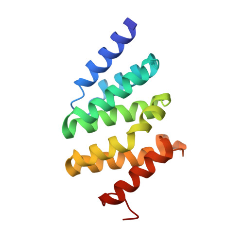
    During transcription, RNA polymerase (Pol) II synthesizes eukaryotic messenger RNA. Transcription is coupled to RNA processing by the carboxy-terminal domain (CTD) of Pol II, which consists of up to 52 repeats of the sequence Tyr 1-Ser 2-Pro 3-Thr 4-Ser 5-Pro 6-Ser 7 (refs 1, 2). After phosphorylation, the CTD binds tightly to a conserved CTD-interacting domain (CID) present in the proteins Pcf11 and Nrd1, which are essential and evolutionarily conserved factors for polyadenylation-dependent and -independent 3'-RNA processing, respectively. Here we describe the structure of a Ser 2-phosphorylated CTD peptide bound to the CID domain of Pcf11. The CTD motif Ser 2-Pro 3-Thr 4-Ser 5 forms a beta-turn that binds to a conserved groove in the CID domain. The Ser 2 phosphate group does not make direct contact with the CID domain, but may be recognized indirectly because it stabilizes the beta-turn with an additional hydrogen bond. Iteration of the peptide structure results in a compact beta-spiral model of the CTD. The model suggests that, during the mRNA transcription-processing cycle, compact spiral regions in the CTD are unravelled and regenerated in a phosphorylation-dependent manner.


  • Organizational Affiliation
    • Department of Chemistry and Biochemistry, Gene Center, University of Munich, 81377 Munich, Germany.

Macromolecule Content 

  • Total Structure Weight: 49.87 kDa 
  • Atom Count: 3,567 
  • Modeled Residue Count: 426 
  • Deposited Residue Count: 432 
  • Unique protein chains: 1

Macromolecules

Find similar proteins by:|  3D Structure
Entity ID: 1
MoleculeChains  Sequence LengthOrganismDetailsImage
PCF11 protein
A, B, C
144Saccharomyces cerevisiaeMutation(s): 0 
Gene Names: PCF11YDR228CYD9934.13C
UniProt
Find proteins for P39081 (Saccharomyces cerevisiae (strain ATCC 204508 / S288c))
Explore P39081 
Go to UniProtKB:  P39081
Entity Groups
Sequence Clusters30% Identity50% Identity70% Identity90% Identity95% Identity100% Identity
UniProt GroupP39081
Sequence Annotations
Expand
Reference Sequence

Experimental Data & Validation

Experimental Data

  • Method: X-RAY DIFFRACTION
  • Resolution: 2.10 Å
  • R-Value Free:  0.252 (Depositor), 0.253 (DCC) 
  • R-Value Work:  0.220 (Depositor), 0.220 (DCC) 
  • R-Value Observed: 0.220 (Depositor) 
Space Group: P 21 21 21
Unit Cell:
Length ( Å )Angle ( ˚ )
a = 56.73α = 90
b = 67.67β = 90
c = 135.637γ = 90
Software Package:
Software NamePurpose
DENZOdata reduction
SCALEPACKdata scaling
SnBphasing
CNSrefinement

Structure Validation

View Full Validation Report


Entry History 

Deposition Data

Revision History  (Full details and data files)

  • Version 1.0: 2004-07-13
    Type: Initial release
  • Version 1.1: 2008-04-30
    Changes: Version format compliance
  • Version 1.2: 2011-07-13
    Changes: Version format compliance
  • Version 1.3: 2017-10-11
    Changes: Refinement description
  • Version 1.4: 2024-02-14
    Changes: Data collection, Database references
  • Version 1.5: 2024-04-03
    Changes: Refinement description