1RHB | pdb_00001rhb

WATER DEPENDENT DOMAIN MOTION AND FLEXIBILITY IN RIBONUCLEASE A AND THE INVARIANT FEATURES IN ITS HYDRATION SHELL. AN X-RAY STUDY OF TWO LOW HUMIDITY CRYSTAL FORMS OF THE ENZYME


Experimental Data Snapshot

  • Method: X-RAY DIFFRACTION
  • Resolution: 1.50 Å
  • R-Value Work: 
    0.158 (DCC) 
  • R-Value Observed: 
    0.173 (Depositor) 

wwPDB Validation 3D Report Full Report

Validation slider image for 1RHB

This is version 1.4 of the entry. See complete history

Literature

Water-dependent domain motion and flexibility in ribonuclease A and the invariant features in its hydration shell. An X-ray study of two low-humidity crystal forms of the enzyme.

Kishan, R.V.Chandra, N.R.Sudarsanakumar, C.Suguna, K.Vijayan, M.

(1995) Acta Crystallogr D Biol Crystallogr 51: 703-710

  • DOI: https://doi.org/10.1107/S0907444994014794
  • Primary Citation Related Structures: 
    1RHA, 1RHB

  • PubMed Abstract: 

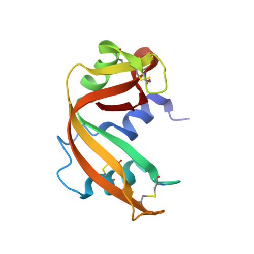
    The crystal structures of 88 and 79% relative humidity forms of ribonuclease A, resulting from water-mediated transformations, have been refined employing the restrained least-squares method using X-ray data collected on an area detector to R = 0.173 for 15 326 observed reflections in the 10-1.5 A resolution shell and R = 0.176 for 8534 observed reflections in the 10-1.8 A shell, respectively. The comparison of these structures with those of the native, the phosphate-bound and the sulfate-bound forms demonstrates that the mobility of the ribonuclease A molecule involves hinge-bending movement of the two domains and local flexibility within them, particularly at the termini of regular secondary structures and in loops. The comparison also leads to the identification of 31 invariant water molecules in the hydration shell of the enzyme, many of which are involved in holding different parts of the molecule together and in stabilizing local structure. The conformational changes that accompany the partial removal of the surrounding water, particularly those observed in the 79% form, could be similar to those that occur during enzyme action.


  • Organizational Affiliation
    • Molecular Biophysics Unit, Indian Institute of Science, Bangalore, India.

Macromolecule Content 

  • Total Structure Weight: 13.71 kDa 
  • Atom Count: 1,107 
  • Modeled Residue Count: 124 
  • Deposited Residue Count: 124 
  • Unique protein chains: 1

Macromolecules

Find similar proteins by:|  3D Structure
Entity ID: 1
MoleculeChains  Sequence LengthOrganismDetailsImage
RIBONUCLEASE A124Bos taurusMutation(s): 0 
EC: 3.1.27.5 (PDB Primary Data), 4.6.1.18 (UniProt)
UniProt
Find proteins for P61823 (Bos taurus)
Explore P61823 
Go to UniProtKB:  P61823
Entity Groups
Sequence Clusters30% Identity50% Identity70% Identity90% Identity95% Identity100% Identity
UniProt GroupP61823
Sequence Annotations
Expand
Reference Sequence

Experimental Data & Validation

Experimental Data

  • Method: X-RAY DIFFRACTION
  • Resolution: 1.50 Å
  • R-Value Work:  0.158 (DCC) 
  • R-Value Observed: 0.173 (Depositor) 
Space Group: P 1 21 1
Unit Cell:
Length ( Å )Angle ( ˚ )
a = 29.74α = 90
b = 38.25β = 112.66
c = 52.4γ = 90
Software Package:
Software NamePurpose
PROLSQrefinement

Structure Validation

View Full Validation Report


Entry History 

Deposition Data

Revision History  (Full details and data files)

  • Version 1.0: 1995-02-27
    Type: Initial release
  • Version 1.1: 2008-03-24
    Changes: Version format compliance
  • Version 1.2: 2011-07-13
    Changes: Version format compliance
  • Version 1.3: 2024-06-05
    Changes: Data collection, Database references, Other
  • Version 1.4: 2024-10-23
    Changes: Structure summary