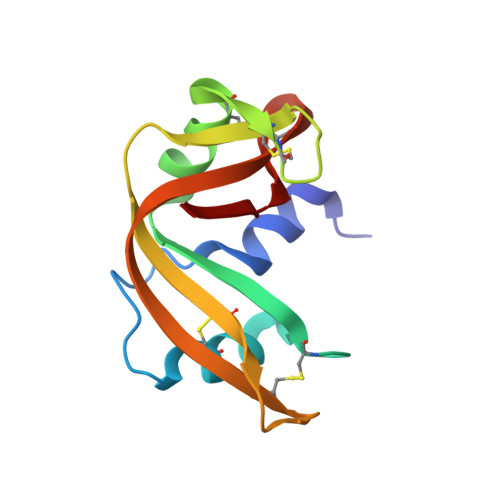
Water-dependent domain motion and flexibility in ribonuclease A and the invariant features in its hydration shell. An X-ray study of two low-humidity crystal forms of the enzyme.
Kishan, R.V., Chandra, N.R., Sudarsanakumar, C., Suguna, K., Vijayan, M.(1995) Acta Crystallogr D Biol Crystallogr 51: 703-710
- PubMed: 15299799
- DOI: https://doi.org/10.1107/S0907444994014794
- Primary Citation of Related Structures:
1RHA, 1RHB - PubMed Abstract:
The crystal structures of 88 and 79% relative humidity forms of ribonuclease A, resulting from water-mediated transformations, have been refined employing the restrained least-squares method using X-ray data collected on an area detector to R = 0.173 for 15 326 observed reflections in the 10-1.5 A resolution shell and R = 0.176 for 8534 observed reflections in the 10-1.8 A shell, respectively. The comparison of these structures with those of the native, the phosphate-bound and the sulfate-bound forms demonstrates that the mobility of the ribonuclease A molecule involves hinge-bending movement of the two domains and local flexibility within them, particularly at the termini of regular secondary structures and in loops. The comparison also leads to the identification of 31 invariant water molecules in the hydration shell of the enzyme, many of which are involved in holding different parts of the molecule together and in stabilizing local structure. The conformational changes that accompany the partial removal of the surrounding water, particularly those observed in the 79% form, could be similar to those that occur during enzyme action.
Organizational Affiliation:
Molecular Biophysics Unit, Indian Institute of Science, Bangalore, India.


