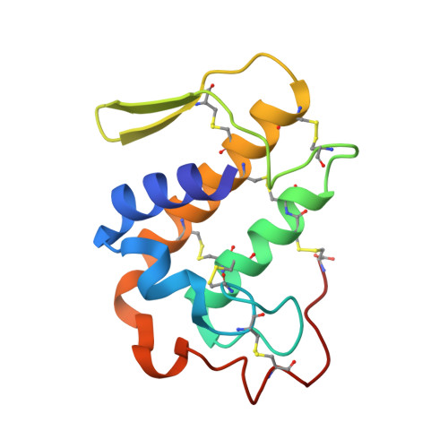
The X-ray structure of a snake venom Gln48 phospholipase A2 at 1.9A resolution reveals anion-binding sites.
Georgieva, D.N., Perbandt, M., Rypniewski, W., Hristov, K., Genov, N., Betzel, C.(2004) Biochem Biophys Res Commun 316: 33-38
- PubMed: 15003507 Search on PubMed
- DOI: https://doi.org/10.1016/j.bbrc.2004.01.174
- Primary Citation Related Structures:
1Q5T - PubMed Abstract:
Phospholipase A2 is an "interfacial" enzyme and its binding to negatively charged surfaces is an important step during catalysis. The Gln48 phospholipase A2 from the venom of Vipera ammodytes meridionalis plays the role of chaperone and directs a toxic His48 PLA2 onto its acceptor. In the venom the two phospholipases A2 exist as a postsynaptic neurotoxic complex, Vipoxin. The X-ray structure of Gln48 PLA2, complexed to sulphate ions, which mimic the negatively charged groups of anionic membranes, has been determined by the molecular replacement method and refined to 1.9A resolution. The protein forms a homodimer stabilized by ionic, hydrophobic, and hydrogen-bond interactions. The structure reveals two anion-binding sites per subunit. These sites are probably involved in interactions with the negatively charged membrane surface and, in this way, in the "targeting" of the toxic component to the receptors of the postsynaptic membranes. In the absence of the chaperone subunit the toxin changes the target of the physiological attack. A comparison of the homodimeric Gln48 PLA2 structure with that of the heterodimeric Vipoxin reveals differences in regions involved in the pharmacological activity of the toxin. This fact, except the active site histidine substitution, can explain the absence of toxicity in the Gln48 protein in comparison to the His48 phospholipase A2.
- Universitätsklinikum Hamburg-Eppendorf, Zentrum für Experimentelle Medizin, Institut für Biochemie und Molekularbiologie I, c/o DESY, Notkestrasse 85, Geb. 22a, Hamburg 22603, Germany.
Organizational Affiliation: