1O0A | pdb_00001o0a

BACTERIORHODOPSIN L INTERMEDIATE AT 1.62 A RESOLUTION


Experimental Data Snapshot

  • Method: X-RAY DIFFRACTION
  • Resolution: 1.62 Å
  • R-Value Free: 
    0.211 (Depositor) 
  • R-Value Work: 
    0.154 (Depositor) 

Starting Model: experimental
View more details

wwPDB Validation 3D Report Full Report

Validation slider image for 1O0A

This is version 1.4 of the entry. See complete history

Literature

Mechanism of proton transport in bacteriorhodopsin from crystallographic structures of the K, L, M1, M2, and M2' intermediates of the photocycle.

Lanyi, J.K.Schobert, B.

(2003) J Mol Biology 328: 439-450

  • DOI: https://doi.org/10.1016/s0022-2836(03)00263-8
  • Primary Citation Related Structures: 
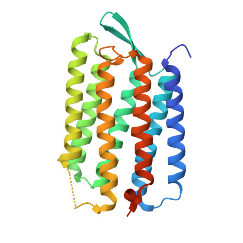
    1O0A

  • PubMed Abstract: 

    We produced the L intermediate of the photocycle in a bacteriorhodopsin crystal in photo-stationary state at 170 K with red laser illumination at 60% occupancy, and determined its structure to 1.62 A resolution. With this model, high-resolution structural information is available for the initial bacteriorhodopsin, as well as the first five states in the transport cycle. These states involve photo-isomerization of the retinal and its initial configurational changes, deprotonation of the retinal Schiff base and the coupled release of a proton to the extracellular membrane surface, and the switch event that allows reprotonation of the Schiff base from the cytoplasmic side. The six structural models describe the transformations of the retinal and its interaction with water 402, Asp85, and Asp212 in atomic detail, as well as the displacements of functional residues farther from the Schiff base. The changes provide rationales for how relaxation of the distorted retinal causes movements of water and protein atoms that result in vectorial proton transfers to and from the Schiff base.


  • Organizational Affiliation
    • Department of Physiology and Biophysics, University of California, 346D Medical Science I, Irvine, CA 92697, USA. jlanyi@orion.oac.uci.edu

Macromolecule Content 

  • Total Structure Weight: 35.9 kDa 
  • Atom Count: 2,072 
  • Modeled Residue Count: 222 
  • Deposited Residue Count: 249 
  • Unique protein chains: 1

Macromolecules

Find similar proteins by:|  3D Structure
Entity ID: 1
MoleculeChains  Sequence LengthOrganismDetailsImage
Bacteriorhodopsin249Halobacterium salinarumMutation(s): 0 
Gene Names: BOP
Membrane Entity: Yes 
UniProt
Find proteins for P02945 (Halobacterium salinarum (strain ATCC 700922 / JCM 11081 / NRC-1))
Explore P02945 
Go to UniProtKB:  P02945
Entity Groups
Sequence Clusters30% Identity50% Identity70% Identity90% Identity95% Identity100% Identity
UniProt GroupP02945
Sequence Annotations
Expand
Reference Sequence

Small Molecules

Ligands 3 Unique
IDChains Name / Formula / InChI Key2D Diagram3D Interactions
LI1

Query on LI1


Download:Ideal Coordinates CCD File
C [auth A]
D [auth A]
E [auth A]
F [auth A]
G [auth A]
C [auth A],
D [auth A],
E [auth A],
F [auth A],
G [auth A],
H [auth A],
I [auth A],
J [auth A],
K [auth A],
L [auth A],
M [auth A],
N [auth A],
O [auth A]
1-[2,6,10.14-TETRAMETHYL-HEXADECAN-16-YL]-2-[2,10,14-TRIMETHYLHEXADECAN-16-YL]GLYCEROL
C42 H86 O3
YERVUJAKCNBGCR-BIHSMRAKSA-N
SQU

Query on SQU


Download:Ideal Coordinates CCD File
P [auth A]2,10,23-TRIMETHYL-TETRACOSANE
C27 H56
ZADHKSJXSZBQFB-HHHXNRCGSA-N
RET

Query on RET


Download:Ideal Coordinates CCD File
B [auth A]RETINAL
C20 H28 O
NCYCYZXNIZJOKI-OVSJKPMPSA-N

Experimental Data & Validation

Experimental Data

  • Method: X-RAY DIFFRACTION
  • Resolution: 1.62 Å
  • R-Value Free:  0.211 (Depositor) 
  • R-Value Work:  0.154 (Depositor) 
Space Group: P 63
Unit Cell:
Length ( Å )Angle ( ˚ )
a = 60.801α = 90
b = 60.801β = 90
c = 107.778γ = 120
Software Package:
Software NamePurpose
HKL-2000data collection
HKL-2000data reduction
SHELXmodel building
SHELXL-97refinement
HKL-2000data reduction
HKL-2000data scaling
SHELXphasing

Structure Validation

View Full Validation Report


Entry History 

Deposition Data

  • Released Date: 2003-04-22 
  • Deposition Author(s): Lanyi, J.K.

Revision History  (Full details and data files)

  • Version 1.0: 2003-04-22
    Type: Initial release
  • Version 1.1: 2008-04-29
    Changes: Version format compliance
  • Version 1.2: 2011-07-13
    Changes: Derived calculations, Version format compliance
  • Version 1.3: 2023-08-16
    Changes: Data collection, Database references, Derived calculations, Refinement description
  • Version 1.4: 2024-11-20
    Changes: Structure summary