1MBS | pdb_00001mbs

X-RAY CRYSTALLOGRAPHIC STUDIES OF SEAL MYOGLOBIN. THE MOLECULE AT 2.5 ANGSTROMS RESOLUTION


Experimental Data Snapshot

  • Method: X-RAY DIFFRACTION
  • Resolution: 2.50 Å

wwPDB Validation 3D Report Full Report

Validation slider image for 1MBS

This is version 1.3 of the entry. See complete history

Literature

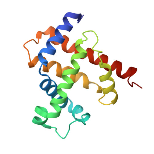
X-ray crystallographic studies of seal myoglobin. The molecule at 2.5 A resolution.

Scouloudi, H.Baker, E.N.

(1978) J Mol Biology 126: 637-660

Macromolecule Content 

  • Total Structure Weight: 17.95 kDa 
  • Atom Count: 1,266 
  • Modeled Residue Count: 153 
  • Deposited Residue Count: 153 
  • Unique protein chains: 1

Macromolecules

Find similar proteins by:|  3D Structure
Entity ID: 1
MoleculeChains  Sequence LengthOrganismDetailsImage
MYOGLOBIN153Phoca vitulinaMutation(s): 0 
EC: 1.11.1 (UniProt), 1.7 (UniProt)
UniProt
Find proteins for P68081 (Halichoerus grypus)
Explore P68081 
Go to UniProtKB:  P68081
Entity Groups
Sequence Clusters30% Identity50% Identity70% Identity90% Identity95% Identity100% Identity
UniProt GroupP68081
Sequence Annotations
Expand
Reference Sequence

Small Molecules

Ligands 1 Unique
IDChains Name / Formula / InChI Key2D Diagram3D Interactions
HEM

Query on HEM


Download:Ideal Coordinates CCD File
B [auth A]PROTOPORPHYRIN IX CONTAINING FE
C34 H32 Fe N4 O4
KABFMIBPWCXCRK-RGGAHWMASA-L

Experimental Data & Validation

Experimental Data

  • Method: X-RAY DIFFRACTION
  • Resolution: 2.50 Å
Space Group: A 1 2 1
Unit Cell:
Length ( Å )Angle ( ˚ )
a = 57.81α = 90
b = 29.7β = 101.99
c = 106.09γ = 90

Structure Validation

View Full Validation Report


Entry History 

Deposition Data

Revision History  (Full details and data files)

  • Version 1.0: 1979-05-15
    Type: Initial release
  • Version 1.1: 2008-03-24
    Changes: Version format compliance
  • Version 1.2: 2011-07-13
    Changes: Version format compliance
  • Version 1.3: 2024-02-14
    Changes: Data collection, Database references, Derived calculations, Other