1M8P | pdb_00001m8p

Crystal Structure of P. chrysogenum ATP Sulfurylase in the T-state


Experimental Data Snapshot

  • Method: X-RAY DIFFRACTION
  • Resolution: 2.60 Å
  • R-Value Free: 
    0.272 (Depositor), 0.273 (DCC) 
  • R-Value Work: 
    0.218 (Depositor), 0.218 (DCC) 
  • R-Value Observed: 
    0.218 (Depositor) 

Starting Model: experimental
View more details

wwPDB Validation 3D Report Full Report

Validation slider image for 1M8P

Ligand Structure Quality Assessment 


This is version 1.3 of the entry. See complete history

Literature

Allosteric Inhibition via R-State Destabilization in ATP Sulfurylase from Penicillium chrysogenum

MacRae, I.J.Segel, I.H.Fisher, A.J.

(2002) Nat Struct Biol 9: 945-949

  • DOI: https://doi.org/10.1038/nsb868
  • Primary Citation Related Structures: 
    1M8P

  • PubMed Abstract: 

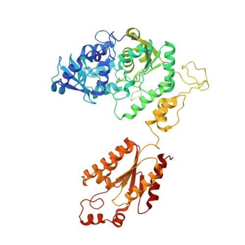
    The structure of the cooperative hexameric enzyme ATP sulfurylase from Penicillium chrysogenum bound to its allosteric inhibitor, 3'-phosphoadenosine-5'-phosphosulfate (PAPS), was determined to 2.6 A resolution. This structure represents the low substrate-affinity T-state conformation of the enzyme. Comparison with the high substrate-affinity R-state structure reveals that a large rotational rearrangement of domains occurs as a result of the R-to-T transition. The rearrangement is accompanied by the 17 A movement of a 10-residue loop out of the active site region, resulting in an open, product release-like structure of the catalytic domain. Binding of PAPS is proposed to induce the allosteric transition by destabilizing an R-state-specific salt linkage between Asp 111 in an N-terminal domain of one subunit and Arg 515 in the allosteric domain of a trans-triad subunit. Disrupting this salt linkage by site-directed mutagenesis induces cooperative inhibition behavior in the absence of an allosteric effector, confirming the role of these two residues.


  • Organizational Affiliation
    • Section of Molecular and Cellular Biology, University of California, One Shields Avenue, Davis, California 95616, USA.

Macromolecule Content 

  • Total Structure Weight: 193.71 kDa 
  • Atom Count: 14,054 
  • Modeled Residue Count: 1,719 
  • Deposited Residue Count: 1,719 
  • Unique protein chains: 1

Macromolecules

Find similar proteins by:|  3D Structure
Entity ID: 1
MoleculeChains  Sequence LengthOrganismDetailsImage
sulfate adenylyltransferase
A, B, C
573Penicillium chrysogenumMutation(s): 0 
EC: 2.7.7.4
UniProt
Find proteins for Q12650 (Penicillium chrysogenum)
Explore Q12650 
Go to UniProtKB:  Q12650
Entity Groups
Sequence Clusters30% Identity50% Identity70% Identity90% Identity95% Identity100% Identity
UniProt GroupQ12650
Sequence Annotations
Expand
Reference Sequence

Experimental Data & Validation

Experimental Data

  • Method: X-RAY DIFFRACTION
  • Resolution: 2.60 Å
  • R-Value Free:  0.272 (Depositor), 0.273 (DCC) 
  • R-Value Work:  0.218 (Depositor), 0.218 (DCC) 
  • R-Value Observed: 0.218 (Depositor) 
Space Group: P 31 2 1
Unit Cell:
Length ( Å )Angle ( ˚ )
a = 135.449α = 90
b = 135.449β = 90
c = 234.587γ = 120
Software Package:
Software NamePurpose
DENZOdata reduction
SCALEPACKdata scaling
CNSrefinement
CNSphasing

Structure Validation

View Full Validation Report


Ligand Structure Quality Assessment 


Entry History 

Deposition Data

Revision History  (Full details and data files)

  • Version 1.0: 2002-11-27
    Type: Initial release
  • Version 1.1: 2008-04-01
    Changes: Version format compliance
  • Version 1.2: 2011-07-13
    Changes: Derived calculations, Version format compliance
  • Version 1.3: 2024-02-14
    Changes: Data collection, Database references, Derived calculations, Refinement description