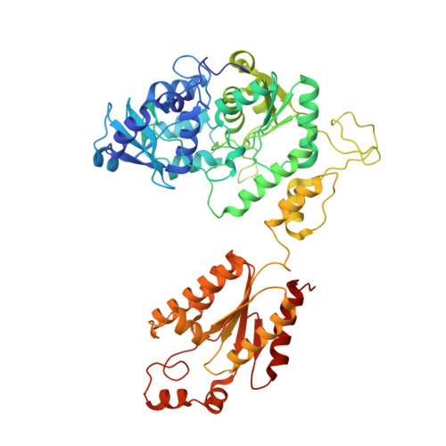
1M8P

Crystal Structure of P. chrysogenum ATP Sulfurylase in the T-state


Experimental Data Snapshot

  • Method: X-RAY DIFFRACTION
  • Resolution: 2.60 Å
  • R-Value Free: 0.272 
  • R-Value Work: 0.218 
  • R-Value Observed: 0.218 

Starting Model: experimental
View more details

wwPDB Validation   3D Report Full Report


Ligand Structure Quality Assessment 


This is version 1.3 of the entry. See complete history


Literature

Allosteric Inhibition via R-State Destabilization in ATP Sulfurylase from Penicillium chrysogenum

MacRae, I.J.Segel, I.H.Fisher, A.J.

(2002) Nat Struct Biol 9: 945-949

  • DOI: https://doi.org/10.1038/nsb868
  • Primary Citation of Related Structures:  
    1M8P

  • PubMed Abstract: 

    The structure of the cooperative hexameric enzyme ATP sulfurylase from Penicillium chrysogenum bound to its allosteric inhibitor, 3'-phosphoadenosine-5'-phosphosulfate (PAPS), was determined to 2.6 A resolution. This structure represents the low substrate-affinity T-state conformation of the enzyme. Comparison with the high substrate-affinity R-state structure reveals that a large rotational rearrangement of domains occurs as a result of the R-to-T transition. The rearrangement is accompanied by the 17 A movement of a 10-residue loop out of the active site region, resulting in an open, product release-like structure of the catalytic domain. Binding of PAPS is proposed to induce the allosteric transition by destabilizing an R-state-specific salt linkage between Asp 111 in an N-terminal domain of one subunit and Arg 515 in the allosteric domain of a trans-triad subunit. Disrupting this salt linkage by site-directed mutagenesis induces cooperative inhibition behavior in the absence of an allosteric effector, confirming the role of these two residues.


  • Organizational Affiliation

    Section of Molecular and Cellular Biology, University of California, One Shields Avenue, Davis, California 95616, USA.


Macromolecules
Find similar proteins by:  (by identity cutoff)  |  3D Structure
Entity ID: 1
MoleculeChains Sequence LengthOrganismDetailsImage
sulfate adenylyltransferase
A, B, C
573Penicillium chrysogenumMutation(s): 0 
EC: 2.7.7.4
UniProt
Find proteins for Q12650 (Penicillium chrysogenum)
Explore Q12650 
Go to UniProtKB:  Q12650
Entity Groups  
Sequence Clusters30% Identity50% Identity70% Identity90% Identity95% Identity100% Identity
UniProt GroupQ12650
Sequence Annotations
Expand
  • Reference Sequence
Experimental Data & Validation

Experimental Data

  • Method: X-RAY DIFFRACTION
  • Resolution: 2.60 Å
  • R-Value Free: 0.272 
  • R-Value Work: 0.218 
  • R-Value Observed: 0.218 
  • Space Group: P 31 2 1
Unit Cell:
Length ( Å )Angle ( ˚ )
a = 135.449α = 90
b = 135.449β = 90
c = 234.587γ = 120
Software Package:
Software NamePurpose
DENZOdata reduction
SCALEPACKdata scaling
CNSrefinement
CNSphasing

Structure Validation

View Full Validation Report


Ligand Structure Quality Assessment 


Entry History 

Deposition Data

Revision History  (Full details and data files)

  • Version 1.0: 2002-11-27
    Type: Initial release
  • Version 1.1: 2008-04-01
    Changes: Version format compliance
  • Version 1.2: 2011-07-13
    Changes: Derived calculations, Version format compliance
  • Version 1.3: 2024-02-14
    Changes: Data collection, Database references, Derived calculations, Refinement description