1M10 | pdb_00001m10

Crystal structure of the complex of Glycoprotein Ib alpha and the von Willebrand Factor A1 Domain


Experimental Data Snapshot

  • Method: X-RAY DIFFRACTION
  • Resolution: 3.10 Å
  • R-Value Free: 
    0.296 (Depositor), 0.282 (DCC) 
  • R-Value Work: 
    0.234 (Depositor), 0.230 (DCC) 
  • R-Value Observed: 
    0.237 (Depositor) 

Starting Models: experimental
View more details

wwPDB Validation 3D Report Full Report

Validation slider image for 1M10

This is version 1.5 of the entry. See complete history

Literature

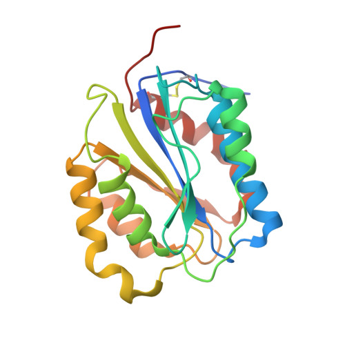
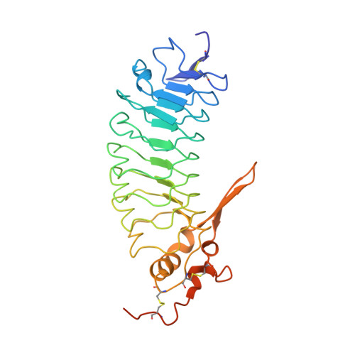
Structures of glycoprotein Ibalpha and its complex with von Willebrand factor A1 domain.

Huizinga, E.G.Tsuji, S.Romijn, R.A.Schiphorst, M.E.de Groot, P.G.Sixma, J.J.Gros, P.

(2002) Science 297: 1176-1179

  • DOI: https://doi.org/10.1126/science.107355
  • Primary Citation Related Structures: 
    1M0Z, 1M10

  • PubMed Abstract: 

    Transient interactions of platelet-receptor glycoprotein Ibalpha (GpIbalpha) and the plasma protein von Willebrand factor (VWF) reduce platelet velocity at sites of vascular damage and play a role in haemostasis and thrombosis. Here we present structures of the GpIbalpha amino-terminal domain and its complex with the VWF domain A1. In the complex, GpIbalpha wraps around one side of A1, providing two contact areas bridged by an area of solvated charge interaction. The structures explain the effects of gain-of-function mutations related to bleeding disorders and provide a model for shear-induced activation. These detailed insights into the initial interactions in platelet adhesion are relevant to the development of antithrombotic drugs.


  • Organizational Affiliation
    • Department of Crystal and Structural Chemistry, Bijvoet Center for Biomolecular Research, Utrecht University, Padualaan 8, 3584 CH Utrecht, Netherlands. e.g.huizinga@chem.uu.nl

Macromolecule Content 

  • Total Structure Weight: 56.07 kDa 
  • Atom Count: 3,688 
  • Modeled Residue Count: 466 
  • Deposited Residue Count: 498 
  • Unique protein chains: 2

Macromolecules

Find similar proteins by:|  3D Structure
Entity ID: 1
MoleculeChains  Sequence LengthOrganismDetailsImage
von Willebrand Factor208Homo sapiensMutation(s): 1 
Gene Names: VWF
UniProt & NIH Common Fund Data Resources
Find proteins for P04275 (Homo sapiens)
Explore P04275 
Go to UniProtKB:  P04275
PHAROS:  P04275
GTEx:  ENSG00000110799 
Entity Groups
Sequence Clusters30% Identity50% Identity70% Identity90% Identity95% Identity100% Identity
UniProt GroupP04275
Sequence Annotations
Expand
Reference Sequence
Find similar proteins by:|  3D Structure
Entity ID: 2
MoleculeChains  Sequence LengthOrganismDetailsImage
Glycoprotein Ib alpha290Homo sapiensMutation(s): 3 
Gene Names: GP1BA
UniProt & NIH Common Fund Data Resources
Find proteins for P07359 (Homo sapiens)
Explore P07359 
Go to UniProtKB:  P07359
PHAROS:  P07359
GTEx:  ENSG00000185245 
Entity Groups
Sequence Clusters30% Identity50% Identity70% Identity90% Identity95% Identity100% Identity
UniProt GroupP07359
Sequence Annotations
Expand
Reference Sequence

Experimental Data & Validation

Experimental Data

  • Method: X-RAY DIFFRACTION
  • Resolution: 3.10 Å
  • R-Value Free:  0.296 (Depositor), 0.282 (DCC) 
  • R-Value Work:  0.234 (Depositor), 0.230 (DCC) 
  • R-Value Observed: 0.237 (Depositor) 
Space Group: P 61
Unit Cell:
Length ( Å )Angle ( ˚ )
a = 89.84α = 90
b = 89.84β = 90
c = 124.63γ = 120
Software Package:
Software NamePurpose
DENZOdata reduction
SCALEPACKdata scaling
CNSrefinement
CNSphasing

Structure Validation

View Full Validation Report


Entry History 

Deposition Data

Revision History  (Full details and data files)

  • Version 1.0: 2002-08-28
    Type: Initial release
  • Version 1.1: 2008-04-28
    Changes: Version format compliance
  • Version 1.2: 2011-07-13
    Changes: Version format compliance
  • Version 1.3: 2021-11-10
    Changes: Data collection, Database references
  • Version 1.4: 2023-10-25
    Changes: Data collection, Refinement description
  • Version 1.5: 2024-10-16
    Changes: Structure summary