1KDG | pdb_00001kdg

Crystal structure of the flavin domain of cellobiose dehydrogenase


Experimental Data Snapshot

  • Method: X-RAY DIFFRACTION
  • Resolution: 1.50 Å
  • R-Value Free: 
    0.169 (Depositor), 0.154 (DCC) 
  • R-Value Work: 
    0.133 (Depositor), 0.147 (DCC) 
  • R-Value Observed: 
    0.135 (Depositor) 

wwPDB Validation 3D Report Full Report

Validation slider image for 1KDG

Ligand Structure Quality Assessment 


This is version 1.6 of the entry. See complete history

Literature

Crystal Structure of the Flavoprotein Domain of the Extracellular Flavocytochrome Cellobiose Dehydrogenase

Hallberg, B.M.Henriksson, G.Pettersson, G.Divne, C.

(2002) J Mol Biology 315: 421-434

  • DOI: https://doi.org/10.1006/jmbi.2001.5246
  • Primary Citation Related Structures: 
    1KDG

  • PubMed Abstract: 

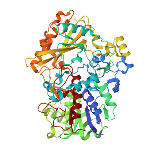
    Cellobiose dehydrogenase (CDH) participates in the degradation of cellulose and lignin. The protein is an extracellular flavocytochrome with a b-type cytochrome domain (CYT(cdh)) connected to a flavodehydrogenase domain (DH(cdh)). DH(cdh) catalyses a two-electron oxidation at the anomeric C1 position of cellobiose to yield cellobiono-1,5-lactone, and the electrons are subsequently transferred from DH(cdh) to an acceptor, either directly or via CYT(cdh). Here, we describe the crystal structure of Phanerochaete chrysosporium DH(cdh) determined at 1.5 A resolution. DH(cdh) belongs to the GMC family of oxidoreductases, which includes glucose oxidase (GOX) and cholesterol oxidase (COX); however, the sequence identity with members of the family is low. The overall fold of DH(cdh) is p-hydroxybenzoate hydroxylase-like and is similar to, but also different from, that of GOX and COX. It is partitioned into an FAD-binding subdomain of alpha/beta type and a substrate-binding subdomain consisting of a seven-stranded beta sheet and six helices. Docking of CYT(cdh) and DH(cdh) suggests that CYT(cdh) covers the active-site entrance in DH(cdh), and that the resulting distance between the cofactors is within acceptable limits for inter-domain electron transfer. Based on docking of the substrate, cellobiose, in the active site of DH(cdh), we propose that the enzyme discriminates against glucose by favouring interaction with the non-reducing end of cellobiose.


  • Organizational Affiliation
    • Department of Cell and Molecular Biology, Structural Biology, Biomedical Centre, Uppsala University, SE-751 24 Uppsala, Sweden.

Macromolecule Content 

  • Total Structure Weight: 121.62 kDa 
  • Atom Count: 9,548 
  • Modeled Residue Count: 1,087 
  • Deposited Residue Count: 1,092 
  • Unique protein chains: 1

Macromolecules

Find similar proteins by:|  3D Structure
Entity ID: 1
MoleculeChains  Sequence LengthOrganismDetailsImage
cellobiose dehydrogenase
A, B
546Phanerodontia chrysosporiumMutation(s): 0 
EC: 1.1.99.18
UniProt
Find proteins for Q01738 (Phanerodontia chrysosporium)
Explore Q01738 
Go to UniProtKB:  Q01738
Entity Groups
Sequence Clusters30% Identity50% Identity70% Identity90% Identity95% Identity100% Identity
UniProt GroupQ01738
Glycosylation
Glycosylation Sites: 5
Sequence Annotations
Expand
Reference Sequence

Small Molecules

Ligands 6 Unique
IDChains Name / Formula / InChI Key2D Diagram3D Interactions
6FA

Query on 6FA


Download:Ideal Coordinates CCD File
M [auth A],
W [auth B]
6-HYDROXY-FLAVIN-ADENINE DINUCLEOTIDE
C27 H33 N9 O16 P2
BJSUUWFQAMLNKU-OKXKTURISA-N
EMT

Query on EMT


Download:Ideal Coordinates CCD File
N [auth A]2-(ETHYLMERCURI-THIO)-BENZOIC ACID
C9 H10 Hg O2 S
HXQVQGWHFRNKMS-UHFFFAOYSA-M
NAG

Query on NAG


Download:Ideal Coordinates CCD File
C [auth A],
D [auth A],
E [auth A],
O [auth B],
P [auth B]
2-acetamido-2-deoxy-beta-D-glucopyranose
C8 H15 N O6
OVRNDRQMDRJTHS-FMDGEEDCSA-N
HG

Query on HG


Download:Ideal Coordinates CCD File
G [auth A]
H [auth A]
I [auth A]
J [auth A]
K [auth A]
G [auth A],
H [auth A],
I [auth A],
J [auth A],
K [auth A],
L [auth A],
T [auth B],
U [auth B],
V [auth B]
MERCURY (II) ION
Hg
BQPIGGFYSBELGY-UHFFFAOYSA-N
MAN

Query on MAN


Download:Ideal Coordinates CCD File
F [auth A],
Q [auth B],
R [auth B],
S [auth B]
alpha-D-mannopyranose
C6 H12 O6
WQZGKKKJIJFFOK-PQMKYFCFSA-N
UNX

Query on UNX


Download:Ideal Coordinates CCD File
X [auth B]UNKNOWN ATOM OR ION
X

Experimental Data & Validation

Experimental Data

  • Method: X-RAY DIFFRACTION
  • Resolution: 1.50 Å
  • R-Value Free:  0.169 (Depositor), 0.154 (DCC) 
  • R-Value Work:  0.133 (Depositor), 0.147 (DCC) 
  • R-Value Observed: 0.135 (Depositor) 
Space Group: H 3
Unit Cell:
Length ( Å )Angle ( ˚ )
a = 185.075α = 90
b = 185.075β = 90
c = 81.034γ = 120
Software Package:
Software NamePurpose
MOSFLMdata reduction
SCALAdata scaling
SOLVEphasing
MLPHAREphasing
REFMACrefinement
CCP4data scaling

Structure Validation

View Full Validation Report


Ligand Structure Quality Assessment 


Entry History 

Deposition Data

Revision History  (Full details and data files)

  • Version 1.0: 2002-11-13
    Type: Initial release
  • Version 1.1: 2008-04-27
    Changes: Version format compliance
  • Version 1.2: 2011-07-13
    Changes: Non-polymer description, Version format compliance
  • Version 1.3: 2012-05-09
    Changes: Structure summary
  • Version 1.4: 2018-03-07
    Changes: Advisory, Data collection
  • Version 1.5: 2020-07-29
    Type: Remediation
    Reason: Carbohydrate remediation
    Changes: Data collection, Derived calculations, Structure summary
  • Version 1.6: 2024-10-16
    Changes: Data collection, Database references, Structure summary