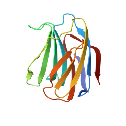
Crystal Structure of a Conger Eel Galectin (Congerin II) at 1.45 A Resolution: Implication for the Accelerated Evolution of a New Ligand-Binding Site Following Gene Duplication
Shirai, T., Matsui, Y., Shionyu-Mitsuyama, C., Yamane, T., Kamiya, H., Ishii, C., Ogawa, T., Muramoto, K.(2002) J Mol Biology 321: 879-889
- PubMed: 12206768
- DOI: https://doi.org/10.1016/s0022-2836(02)00700-3
- Primary Citation of Related Structures:
1IS3, 1IS4, 1IS5, 1IS6 - PubMed Abstract:
The crystal structure of congerin II, a galectin family lectin from conger eel, was determined at 1.45A resolution. The previously determined structure of its isoform, congerin I, had revealed a fold evolution via strand swap; however, the structure of congerin II described here resembles other prototype galectins. A comparison of the two congerin genes with that of several other galectins suggests acceralated evolution of both congerin genes following gene duplication. The presence of a Mes (2-[N-morpholino]ethanesulfonic acid) molecule near the carbohydrate-binding site in the crystal structure points to the possibility of an additional binding site in congerin II. The binding site consists of a group of residues that had been replaced following gene duplication suggesting that the binding site was built under selective pressure. Congerin II may be a protein specialized for biological defense with an affinity for target carbohydrates on parasites' cell surface.
- Department of Biotechnology and Biomaterial Chemistry, Graduate School of Engineering, Nagoya University, Chikusa-Ku, Japan. i45282a@nucc.cc.nagoya-u.ac.jp
Organizational Affiliation:


