Functional Analyses of the Domain Structure in the Holliday Junction Binding Protein Ruva
Nishino, T., Ariyoshi, M., Iwasaki, H., Shinagawa, H., Morikawa, K.(1998) Structure 6: 11-21
- PubMed: 9493263 Search on PubMed
- DOI: https://doi.org/10.1016/s0969-2126(98)00003-3
- Primary Citation Related Structures:
1HJP - PubMed Abstract:
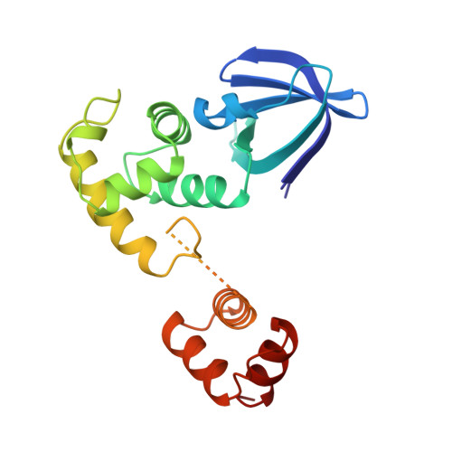
Homologous recombination is crucial for genetic diversity and repairing damaged chromosomes. In Escherichia coli cells, the RuvA, RuvB and RuvC proteins participate in the processing of an important intermediate, the Holliday junction. The RuvA-RuvB protein complex facilitates branch migration of the junction, depending on ATP hydrolysis. The atomic structure of RuvA should enable critical questions to be addressed about its specific interactions with the Holliday junction and the RuvB protein. The crystal structure of RuvA shows the tetrameric molecules with a fourfold axis at the center. Each subunit consists of three distinct domains, some of which contain important secondary structure elements for DNA binding. Together with the detailed structural information, the biochemical assays of various mutant RuvA proteins and domains, isolated by partial proteolysis, allowed us to define the functional roles of these domains in Holliday junction binding and the RuvB interaction. The RuvA molecule is formed by four identical subunits, each with three domains, I, II and III. The locations of the putative DNA-binding motifs define an interface between the DNA and the Holliday junction. Domain III is weakly attached to the core region, comprising domains I and II; the core domains can form a tetramer in the absence of domain III. Functional analyses of the mutant proteins and the partial digestion products, including Holliday junction binding and branch-migration assays, revealed that domain III and the preceding loop are crucial for RuvB binding and branch migration, although this region is not required for the junction-DNA binding.
- Department of Structural Biology, Biomolecular Engineering Research Institute (BERI), Osaka, Japan.
Organizational Affiliation: