1H6U

Internalin H: crystal structure of fused N-terminal domains.


Experimental Data Snapshot

  • Method: X-RAY DIFFRACTION
  • Resolution: 1.80 Å
  • R-Value Free: 0.217 
  • R-Value Work: 0.167 
  • R-Value Observed: 0.170 

Starting Model: experimental
View more details

wwPDB Validation   3D Report Full Report


This is version 1.4 of the entry. See complete history


Literature

Internalins from the human pathogen Listeria monocytogenes combine three distinct folds into a contiguous internalin domain.

Schubert, W.D.Gobel, G.Diepholz, M.Darji, A.Kloer, D.Hain, T.Chakraborty, T.Wehland, J.Domann, E.Heinz, D.W.

(2001) J Mol Biol 312: 783-794

  • DOI: https://doi.org/10.1006/jmbi.2001.4989
  • Primary Citation of Related Structures:  
    1H6T, 1H6U

  • PubMed Abstract: 

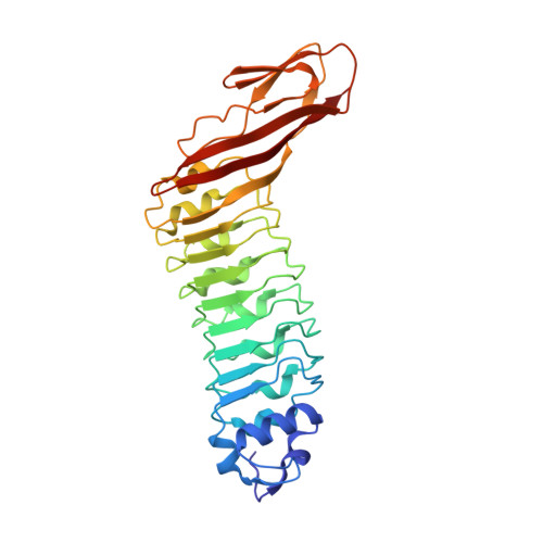
    Listeria monocytogenes is an opportunistic, food-borne human and animal pathogen. Host cell invasion requires the action of the internalins A (InlA) and B (InlB), which are members of a family of listerial cell-surface proteins. Common to these proteins are three distinctive N-terminal domains that have been shown to direct host cell-specific invasion for InlA and InlB. Here, we present the high-resolution crystal structures of these domains present in InlB and InlH, and show that they constitute a single "internalin domain". In this internalin domain, a central LRR region is flanked contiguously by a truncated EF-hand-like cap and an immunoglobulin (Ig)-like fold. The extended beta-sheet, resulting from the distinctive fusion of the LRR and the Ig-like folds, constitutes an adaptable concave interaction surface, which we propose is responsible for the specific recognition of the host cellular binding partners during infection.


  • Organizational Affiliation

    Department of Structural Biology, German Research Center for Biotechnology, Braunschweig.


Macromolecules
Find similar proteins by:  (by identity cutoff)  |  3D Structure
Entity ID: 1
MoleculeChains Sequence LengthOrganismDetailsImage
INTERNALIN H308Listeria monocytogenesMutation(s): 1 
UniProt
Find proteins for Q9ZEY1 (Listeria monocytogenes)
Explore Q9ZEY1 
Go to UniProtKB:  Q9ZEY1
Entity Groups  
Sequence Clusters30% Identity50% Identity70% Identity90% Identity95% Identity100% Identity
UniProt GroupQ9ZEY1
Sequence Annotations
Expand
  • Reference Sequence
Experimental Data & Validation

Experimental Data

  • Method: X-RAY DIFFRACTION
  • Resolution: 1.80 Å
  • R-Value Free: 0.217 
  • R-Value Work: 0.167 
  • R-Value Observed: 0.170 
  • Space Group: I 21 21 21
Unit Cell:
Length ( Å )Angle ( ˚ )
a = 57.481α = 90
b = 100.682β = 90
c = 119.931γ = 90
Software Package:
Software NamePurpose
REFMACrefinement
DENZOdata reduction
SCALEPACKdata scaling
AMoREphasing

Structure Validation

View Full Validation Report


Entry History 

Deposition Data

Revision History  (Full details and data files)

  • Version 1.0: 2001-10-11
    Type: Initial release
  • Version 1.1: 2011-05-08
    Changes: Version format compliance
  • Version 1.2: 2011-07-13
    Changes: Version format compliance
  • Version 1.3: 2019-05-15
    Changes: Data collection, Database references, Experimental preparation, Other
  • Version 1.4: 2023-12-13
    Changes: Data collection, Database references, Refinement description