1CJC

STRUCTURE OF ADRENODOXIN REDUCTASE OF MITOCHONDRIAL P450 SYSTEMS


Experimental Data Snapshot

  • Method: X-RAY DIFFRACTION
  • Resolution: 1.70 Å
  • R-Value Free: 0.223 
  • R-Value Work: 0.188 

wwPDB Validation   3D Report Full Report


This is version 1.5 of the entry. See complete history


Literature

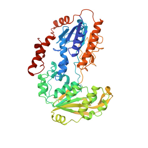
The structure of adrenodoxin reductase of mitochondrial P450 systems: electron transfer for steroid biosynthesis.

Ziegler, G.A.Vonrhein, C.Hanukoglu, I.Schulz, G.E.

(1999) J Mol Biol 289: 981-990

  • DOI: https://doi.org/10.1006/jmbi.1999.2807
  • Primary Citation of Related Structures:  
    1CJC

  • PubMed Abstract: 

    Adrenodoxin reductase is a monomeric 51 kDa flavoenzyme that is involved in the biosynthesis of all steroid hormones. The structure of the native bovine enzyme was determined at 2.8 A resolution, and the structure of the respective recombinant enzyme at 1.7 A resolution. Adrenodoxin reductase receives a two-electron package from NADPH and converts it to two single electrons that are transferred via adrenodoxin to all mitochondrial cytochromes P 450. The structure suggests how the observed flavin semiquinone is stabilized. A striking feature is the asymmetric charge distribution, which most likely controls the approach of the electron carrier adrenodoxin. A model for the interaction is proposed. Adrenodoxin reductase shows clear sequence homology to half a dozen proteins identified in genome analysis projects, but neither sequence nor structural homology to established, functionally related electron transferases. Yet, the structure revealed a relationship to the disulfide oxidoreductases, permitting the assignment of the NADP-binding site.


  • Organizational Affiliation

    Albert-Ludwigs-Universität, Albertstrasse 21, Freiburg im Breisgau, D-79104, Germany.


Macromolecules
Find similar proteins by:  (by identity cutoff)  |  3D Structure
Entity ID: 1
MoleculeChains Sequence LengthOrganismDetailsImage
PROTEIN (ADRENODOXIN REDUCTASE)460Bos taurusMutation(s): 0 
EC: 1.18.1.6
UniProt
Find proteins for P08165 (Bos taurus)
Explore P08165 
Go to UniProtKB:  P08165
Entity Groups  
Sequence Clusters30% Identity50% Identity70% Identity90% Identity95% Identity100% Identity
UniProt GroupP08165
Sequence Annotations
Expand
  • Reference Sequence
Small Molecules
Ligands 1 Unique
IDChains Name / Formula / InChI Key2D Diagram3D Interactions
FAD
Query on FAD

Download Ideal Coordinates CCD File 
B [auth A]FLAVIN-ADENINE DINUCLEOTIDE
C27 H33 N9 O15 P2
VWWQXMAJTJZDQX-UYBVJOGSSA-N
Experimental Data & Validation

Experimental Data

  • Method: X-RAY DIFFRACTION
  • Resolution: 1.70 Å
  • R-Value Free: 0.223 
  • R-Value Work: 0.188 
  • Space Group: P 1 21 1
Unit Cell:
Length ( Å )Angle ( ˚ )
a = 60.81α = 90
b = 62.53β = 106.76
c = 78.37γ = 90
Software Package:
Software NamePurpose
SHARPphasing
REFMACrefinement
MOSFLMdata reduction
CCP4data scaling

Structure Validation

View Full Validation Report


Entry History 

Deposition Data

Revision History  (Full details and data files)

  • Version 1.0: 1999-04-30
    Type: Initial release
  • Version 1.1: 2008-04-26
    Changes: Version format compliance
  • Version 1.2: 2011-07-13
    Changes: Version format compliance
  • Version 1.3: 2013-04-17
    Changes: Structure summary
  • Version 1.4: 2019-11-27
    Changes: Advisory, Data collection, Database references
  • Version 1.5: 2023-12-27
    Changes: Advisory, Data collection, Database references, Derived calculations