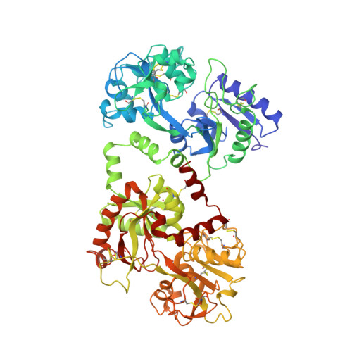
1CE2

STRUCTURE OF DIFERRIC BUFFALO LACTOFERRIN AT 2.5A RESOLUTION


Experimental Data Snapshot

  • Method: X-RAY DIFFRACTION
  • Resolution: 2.50 Å
  • R-Value Free: 0.265 
  • R-Value Work: 0.187 

Starting Model: experimental
View more details

wwPDB Validation   3D Report Full Report


This is version 1.6 of the entry. See complete history


Literature

Structure of buffalo lactoferrin at 2.5 A resolution using crystals grown at 303 K shows different orientations of the N and C lobes.

Karthikeyan, S.Paramasivam, M.Yadav, S.Srinivasan, A.Singh, T.P.

(1999) Acta Crystallogr D Biol Crystallogr 55: 1805-1813

  • DOI: https://doi.org/10.1107/s0907444999010951
  • Primary Citation of Related Structures:  
    1CE2

  • PubMed Abstract: 

    The structure of buffalo lactoferrin has been determined at 303 K. The crystals belong to orthorhombic space group P2(1)2(1)2(1), with unit-cell parameters a = 77.5, b = 91.0, c = 131.5 A and Z = 4. The structure has been refined to an R factor of 0.187. The overall structure of the protein is similar to its structure determined at 277 K in a different crystal form. However, the lobe orientations in the two structures differ by 9.0 degrees, suggesting significant inter-lobe flexibility in this family of proteins. The inter-lobe interactions are predominantly hydrophobic and could act as a cushion for a change in orientation under the influence of external conditions. On the other hand, the domain arrangements are found to be similar in 277 and 303 K crystal structures, with orientations differing by 1.5 and 1.0 degrees in the N and C lobes, respectively. The results of these investigations suggest that the increase in temperature helps in the production of better quality crystals.


  • Organizational Affiliation

    Department of Biophysics, All India Institute of Medical Sciences, New Delhi 110 029, India.


Macromolecules
Find similar proteins by:  (by identity cutoff)  |  3D Structure
Entity ID: 1
MoleculeChains Sequence LengthOrganismDetailsImage
PROTEIN (LACTOFERRIN)689Bubalus bubalisMutation(s): 0 
EC: 3.4.21
UniProt
Find proteins for O77698 (Bubalus bubalis)
Explore O77698 
Go to UniProtKB:  O77698
Entity Groups  
Sequence Clusters30% Identity50% Identity70% Identity90% Identity95% Identity100% Identity
UniProt GroupO77698
Sequence Annotations
Expand
  • Reference Sequence
Experimental Data & Validation

Experimental Data

  • Method: X-RAY DIFFRACTION
  • Resolution: 2.50 Å
  • R-Value Free: 0.265 
  • R-Value Work: 0.187 
  • Space Group: P 21 21 21
Unit Cell:
Length ( Å )Angle ( ˚ )
a = 77.462α = 90
b = 91.035β = 90
c = 131.507γ = 90
Software Package:
Software NamePurpose
DENZOdata reduction
AUTOMARdata reduction
AMoREphasing
X-PLORrefinement

Structure Validation

View Full Validation Report


Entry History 

Deposition Data

Revision History  (Full details and data files)

  • Version 1.0: 1999-03-19
    Type: Initial release
  • Version 1.1: 2007-10-16
    Changes: Version format compliance
  • Version 1.2: 2011-07-13
    Changes: Version format compliance
  • Version 1.3: 2017-10-04
    Changes: Refinement description
  • Version 1.4: 2019-11-06
    Changes: Data collection, Database references
  • Version 1.5: 2023-08-09
    Changes: Data collection, Database references, Derived calculations, Refinement description
  • Version 1.6: 2024-10-30
    Changes: Structure summary