1CE2 | pdb_00001ce2

STRUCTURE OF DIFERRIC BUFFALO LACTOFERRIN AT 2.5A RESOLUTION


Experimental Data Snapshot

  • Method: X-RAY DIFFRACTION
  • Resolution: 2.50 Å
  • R-Value Free: 
    0.265 (Depositor) 
  • R-Value Work: 
    0.187 (Depositor) 

Starting Model: experimental
View more details

wwPDB Validation 3D Report Full Report

Validation slider image for 1CE2

This is version 1.6 of the entry. See complete history

Literature

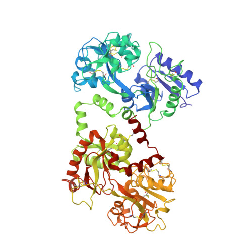
Structure of buffalo lactoferrin at 2.5 A resolution using crystals grown at 303 K shows different orientations of the N and C lobes.

Karthikeyan, S.Paramasivam, M.Yadav, S.Srinivasan, A.Singh, T.P.

(1999) Acta Crystallogr D Biol Crystallogr 55: 1805-1813

  • DOI: https://doi.org/10.1107/s0907444999010951
  • Primary Citation Related Structures: 
    1CE2

  • PubMed Abstract: 

    The structure of buffalo lactoferrin has been determined at 303 K. The crystals belong to orthorhombic space group P2(1)2(1)2(1), with unit-cell parameters a = 77.5, b = 91.0, c = 131.5 A and Z = 4. The structure has been refined to an R factor of 0.187. The overall structure of the protein is similar to its structure determined at 277 K in a different crystal form. However, the lobe orientations in the two structures differ by 9.0 degrees, suggesting significant inter-lobe flexibility in this family of proteins. The inter-lobe interactions are predominantly hydrophobic and could act as a cushion for a change in orientation under the influence of external conditions. On the other hand, the domain arrangements are found to be similar in 277 and 303 K crystal structures, with orientations differing by 1.5 and 1.0 degrees in the N and C lobes, respectively. The results of these investigations suggest that the increase in temperature helps in the production of better quality crystals.


  • Organizational Affiliation
    • Department of Biophysics, All India Institute of Medical Sciences, New Delhi 110 029, India.

Macromolecule Content 

  • Total Structure Weight: 76.08 kDa 
  • Atom Count: 5,415 
  • Modeled Residue Count: 689 
  • Deposited Residue Count: 689 
  • Unique protein chains: 1

Macromolecules

Find similar proteins by:|  3D Structure
Entity ID: 1
MoleculeChains  Sequence LengthOrganismDetailsImage
PROTEIN (LACTOFERRIN)689Bubalus bubalisMutation(s): 0 
EC: 3.4.21
UniProt
Find proteins for O77698 (Bubalus bubalis)
Explore O77698 
Go to UniProtKB:  O77698
Entity Groups
Sequence Clusters30% Identity50% Identity70% Identity90% Identity95% Identity100% Identity
UniProt GroupO77698
Sequence Annotations
Expand
Reference Sequence

Experimental Data & Validation

Experimental Data

  • Method: X-RAY DIFFRACTION
  • Resolution: 2.50 Å
  • R-Value Free:  0.265 (Depositor) 
  • R-Value Work:  0.187 (Depositor) 
Space Group: P 21 21 21
Unit Cell:
Length ( Å )Angle ( ˚ )
a = 77.462α = 90
b = 91.035β = 90
c = 131.507γ = 90
Software Package:
Software NamePurpose
DENZOdata reduction
AUTOMARdata reduction
AMoREphasing
X-PLORrefinement

Structure Validation

View Full Validation Report


Entry History 

Deposition Data

Revision History  (Full details and data files)

  • Version 1.0: 1999-03-19
    Type: Initial release
  • Version 1.1: 2007-10-16
    Changes: Version format compliance
  • Version 1.2: 2011-07-13
    Changes: Version format compliance
  • Version 1.3: 2017-10-04
    Changes: Refinement description
  • Version 1.4: 2019-11-06
    Changes: Data collection, Database references
  • Version 1.5: 2023-08-09
    Changes: Data collection, Database references, Derived calculations, Refinement description
  • Version 1.6: 2024-10-30
    Changes: Structure summary