1ZXX

The crystal structure of phosphofructokinase from Lactobacillus delbrueckii


Experimental Data Snapshot

  • Method: X-RAY DIFFRACTION
  • Resolution: 1.85 Å
  • R-Value Free: 0.251 
  • R-Value Work: 0.222 
  • R-Value Observed: 0.224 

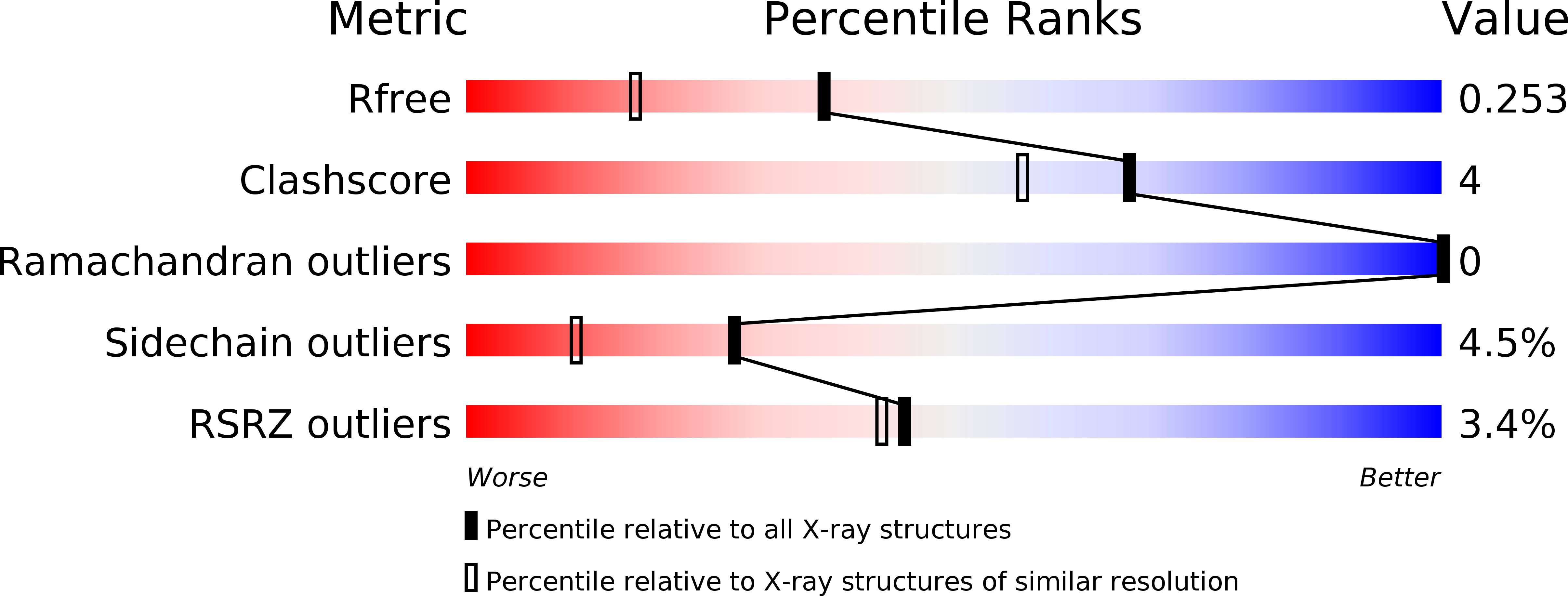
wwPDB Validation   3D Report Full Report


This is version 1.3 of the entry. See complete history


Literature

Kinetic and Structural Characterization of Phosphofructokinase from Lactobacillus bulgaricus.

Paricharttanakul, N.M.Ye, S.Menefee, A.L.Javid-Majd, F.Sacchettini, J.C.Reinhart, G.D.

(2005) Biochemistry 44: 15280-15286

  • DOI: https://doi.org/10.1021/bi051283g
  • Primary Citation of Related Structures:  
    1ZXX

  • PubMed Abstract: 

    Phosphofructokinase from Lactobacillus delbrueckii subspecies bulgaricus (LbPFK) has been reported to be a nonallosteric analogue of phosphofructokinase from Escherichia coli at pH 8.2 [Le Bras et al. (1991) Eur. J. Biochem. 198, 683-687]. A reexamination of the kinetics of this enzyme shows LbPFK to have limited binding affinity toward the allosteric ligands, MgADP and PEP, with dissociation constants of approximately 20 mM for both. Their allosteric effects are observed only at high concentrations of these ligands, with both exhibiting inhibitory effects on substrate binding. No pH dependence was observed for the binding and the influence of MgADP and PEP on the enzyme. To attempt to explain these results, the crystal structure of LbPFK was solved using molecular replacement to 1.86 A resolution. A comparative study of the LbPFK structure with that of phosphofructokinases from E. coli (EcPFK) and Bacillus stearothermophilus (BsPFK) reveals a structure with conserved fold and substrate binding site. The effector binding site, however, shows many differences that could explain the observed decreases in binding affinity for MgADP and PEP in LbPFK as compared to the other two enzymes.


  • Organizational Affiliation

    Department of Biochemistry and Biophysics, Texas A&M University, and the Texas Agricultural Experiment Station, College Station, Texas 77843-2128, USA.


Macromolecules
Find similar proteins by:  (by identity cutoff)  |  3D Structure
Entity ID: 1
MoleculeChains Sequence LengthOrganismDetailsImage
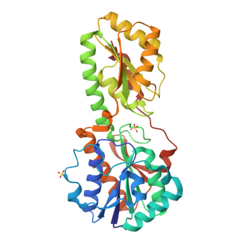
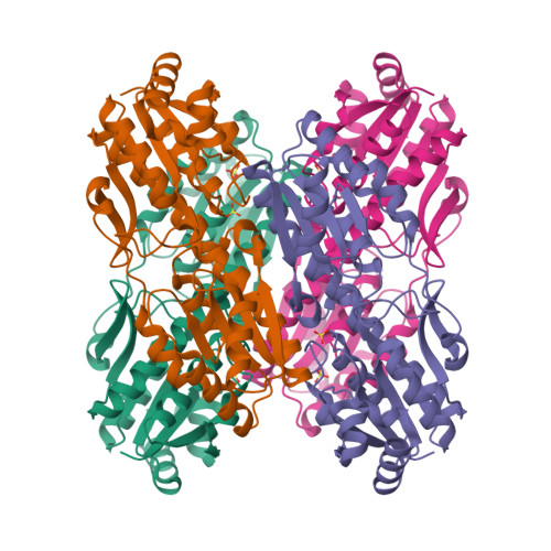
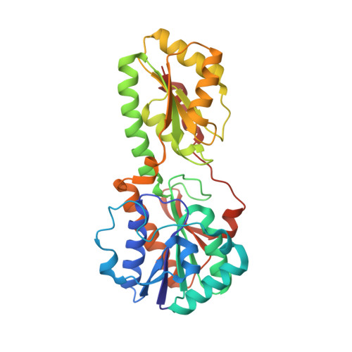
6-phosphofructokinase319Lactobacillus delbrueckii subsp. bulgaricusMutation(s): 0 
Gene Names: pfkA
EC: 2.7.1.11
UniProt
Find proteins for P80019 (Lactobacillus delbrueckii subsp. bulgaricus)
Explore P80019 
Go to UniProtKB:  P80019
Entity Groups  
Sequence Clusters30% Identity50% Identity70% Identity90% Identity95% Identity100% Identity
UniProt GroupP80019
Sequence Annotations
Expand
  • Reference Sequence
Experimental Data & Validation

Experimental Data

  • Method: X-RAY DIFFRACTION
  • Resolution: 1.85 Å
  • R-Value Free: 0.251 
  • R-Value Work: 0.222 
  • R-Value Observed: 0.224 
  • Space Group: P 62 2 2
Unit Cell:
Length ( Å )Angle ( ˚ )
a = 135.014α = 90
b = 135.014β = 90
c = 77.66γ = 120
Software Package:
Software NamePurpose
REFMACrefinement
HKL-2000data reduction
SCALEPACKdata scaling
AMoREphasing

Structure Validation

View Full Validation Report


Entry History 

Deposition Data

Revision History  (Full details and data files)

  • Version 1.0: 2005-11-08
    Type: Initial release
  • Version 1.1: 2008-04-30
    Changes: Version format compliance
  • Version 1.2: 2011-07-13
    Changes: Derived calculations, Version format compliance
  • Version 1.3: 2024-02-14
    Changes: Data collection, Database references, Derived calculations