


The structure of a putative mannosephosphate isomerase from Archaeoglobus fulgidus
Cuff, M.E., Skarina, T., Edwards, A., Savchenko, A., Joachimiak, A.To be published.
Experimental Data Snapshot
Entity ID: 1 | |||||
|---|---|---|---|---|---|
| Molecule | Chains | Sequence Length | Organism | Details | Image |
| mannosephosphate isomerase, putative | 300 | Archaeoglobus fulgidus | Mutation(s): 4 Gene Names: AF0035 EC: 5.3.1.8 | 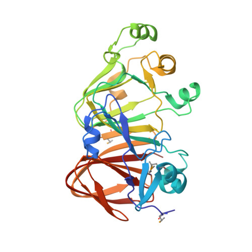 | |
UniProt | |||||
Entity Groups | |||||
| Sequence Clusters | 30% Identity50% Identity70% Identity90% Identity95% Identity100% Identity | ||||
| UniProt Group | O30200 | ||||
Sequence AnnotationsExpand | |||||
Reference Sequence | |||||
| Ligands 4 Unique | |||||
|---|---|---|---|---|---|
| ID | Chains | Name / Formula / InChI Key | 2D Diagram | 3D Interactions | |
| LFR Download:Ideal Coordinates CCD File | B [auth A] | beta-L-fructofuranose C6 H12 O6 RFSUNEUAIZKAJO-AZGQCCRYSA-N |  | ||
| GOL Download:Ideal Coordinates CCD File | K [auth A] | GLYCEROL C3 H8 O3 PEDCQBHIVMGVHV-UHFFFAOYSA-N |  | ||
| EDO Download:Ideal Coordinates CCD File | C [auth A], D [auth A] | 1,2-ETHANEDIOL C2 H6 O2 LYCAIKOWRPUZTN-UHFFFAOYSA-N |  | ||
| ACY Download:Ideal Coordinates CCD File | E [auth A] F [auth A] G [auth A] H [auth A] I [auth A] | ACETIC ACID C2 H4 O2 QTBSBXVTEAMEQO-UHFFFAOYSA-N |  | ||
| Modified Residues 1 Unique | |||||
|---|---|---|---|---|---|
| ID | Chains | Type | Formula | 2D Diagram | Parent |
| MSE Query on MSE | A | L-PEPTIDE LINKING | C5 H11 N O2 Se |  | MET |
| Length ( Å ) | Angle ( ˚ ) |
|---|---|
| a = 160.045 | α = 90 |
| b = 160.045 | β = 90 |
| c = 160.045 | γ = 90 |
| Software Name | Purpose |
|---|---|
| REFMAC | refinement |
| SBC-Collect | data collection |
| HKL-2000 | data scaling |
| HKL-3000 | phasing |
| SHELXD | phasing |
| SHELXE | model building |
| MLPHARE | phasing |
| DM | phasing |
| SOLVE | phasing |
| RESOLVE | phasing |
| O | model building |
| Coot | model building |
| CCP4 | phasing |
| ARP/wARP | model building |


