


Crystal structure of spin labeled T4 Lysozyme (V131R1B)
Fleissner, M.R., Cascio, D., Sawaya, M.R., Hideg, K., Hubbell, W.L.To be published.
Experimental Data Snapshot
Starting Model: experimental
View more details
Entity ID: 1 | |||||
|---|---|---|---|---|---|
| Molecule | Chains | Sequence Length | Organism | Details | Image |
| Lysozyme | 164 | Tequatrovirus T4 | Mutation(s): 3 EC: 3.2.1.17 | 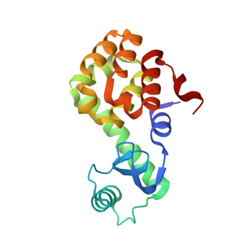 | |
UniProt | |||||
Entity Groups | |||||
| Sequence Clusters | 30% Identity50% Identity70% Identity90% Identity95% Identity100% Identity | ||||
| UniProt Group | P00720 | ||||
Sequence AnnotationsExpand | |||||
Reference Sequence | |||||
| Ligands 4 Unique | |||||
|---|---|---|---|---|---|
| ID | Chains | Name / Formula / InChI Key | 2D Diagram | 3D Interactions | |
| R1B Download:Ideal Coordinates CCD File | B [auth A] | S-[(1-oxyl-2,2,4,5,5-pentamethyl-2,5-dihydro-1H-pyrrol-3-yl)methyl] methanesulfonothioate C11 H20 N O3 S2 MMNQHAFLTLSLNX-UHFFFAOYSA-N |  | ||
| HED Download:Ideal Coordinates CCD File | H [auth A] | 2-HYDROXYETHYL DISULFIDE C4 H10 O2 S2 KYNFOMQIXZUKRK-UHFFFAOYSA-N |  | ||
| AZI Download:Ideal Coordinates CCD File | C [auth A], D [auth A] | AZIDE ION N3 IVRMZWNICZWHMI-UHFFFAOYSA-N |  | ||
| CL Download:Ideal Coordinates CCD File | E [auth A], F [auth A], G [auth A] | CHLORIDE ION Cl VEXZGXHMUGYJMC-UHFFFAOYSA-M |  | ||
| Length ( Å ) | Angle ( ˚ ) |
|---|---|
| a = 59.793 | α = 90 |
| b = 59.793 | β = 90 |
| c = 95.442 | γ = 120 |
| Software Name | Purpose |
|---|---|
| REFMAC | refinement |
| DENZO | data reduction |
| SCALEPACK | data scaling |
| PHASER | phasing |


