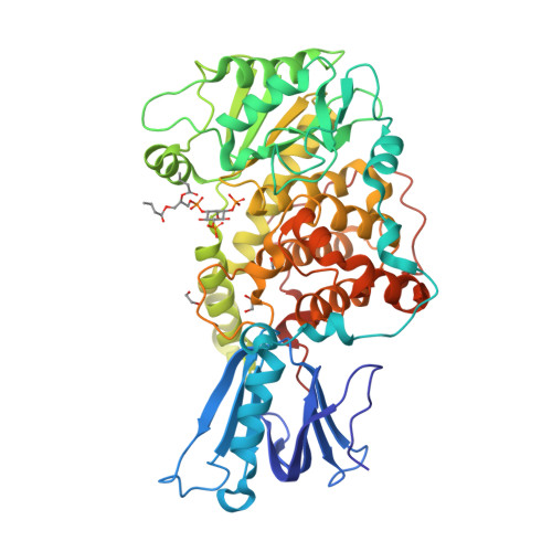
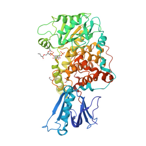
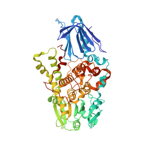
1ZVR

Crystal Structure of MTMR2 in complex with phosphatidylinositol 3,5-bisphosphate


Experimental Data Snapshot

  • Method: X-RAY DIFFRACTION
  • Resolution: 1.98 Å
  • R-Value Free: 0.252 
  • R-Value Work: 0.228 

Starting Model: experimental
View more details

wwPDB Validation   3D Report Full Report


Ligand Structure Quality Assessment 


This is version 2.1 of the entry. See complete history


Literature

Molecular basis for substrate recognition by MTMR2, a myotubularin family phosphoinositide phosphatase

Begley, M.J.Taylor, G.S.Brock, M.A.Ghosh, P.Woods, V.L.Dixon, J.E.

(2006) Proc Natl Acad Sci U S A 103: 927-932

  • DOI: https://doi.org/10.1073/pnas.0510006103
  • Primary Citation of Related Structures:  
    1ZSQ, 1ZVR

  • PubMed Abstract: 

    Myotubularins, a large family of catalytically active and inactive proteins, belong to a unique subgroup of protein tyrosine phosphatases that use inositol phospholipids, rather than phosphoproteins, as physiological substrates. Here, by integrating crystallographic and deuterium-exchange mass spectrometry studies of human myotubularin-related protein-2 (MTMR2) in complex with phosphoinositides, we define the molecular basis for this unique substrate specificity. Phosphoinositide substrates bind in a pocket located on a positively charged face of the protein, suggesting an electrostatic mechanism for membrane targeting. A flexible, hydrophobic helix makes extensive interactions with the diacylglycerol moieties of substrates, explaining the specificity for membrane-bound phosphoinositides. An extensive H-bonding network and charge-charge interactions within the active site pocket determine phosphoinositide headgroup specificity. The conservation of these specificity determinants within the active, but not the inactive, myotubularins provides insight into the functional differences between the active and inactive members.


  • Organizational Affiliation

    Department of Pharmacology, University of California at San Diego, La Jolla, CA 92093, USA.


Macromolecules
Find similar proteins by:  (by identity cutoff)  |  3D Structure
Entity ID: 1
MoleculeChains Sequence LengthOrganismDetailsImage
Myotubularin-related protein 2528Homo sapiensMutation(s): 1 
Gene Names: MTMR2KIAA1073
EC: 3.1.3 (PDB Primary Data), 3.1.3.95 (UniProt)
Membrane Entity: Yes 
UniProt & NIH Common Fund Data Resources
Find proteins for Q13614 (Homo sapiens)
Explore Q13614 
Go to UniProtKB:  Q13614
PHAROS:  Q13614
GTEx:  ENSG00000087053 
Entity Groups  
Sequence Clusters30% Identity50% Identity70% Identity90% Identity95% Identity100% Identity
UniProt GroupQ13614
Sequence Annotations
Expand
  • Reference Sequence
Experimental Data & Validation

Experimental Data

  • Method: X-RAY DIFFRACTION
  • Resolution: 1.98 Å
  • R-Value Free: 0.252 
  • R-Value Work: 0.228 
  • Space Group: P 41 21 2
Unit Cell:
Length ( Å )Angle ( ˚ )
a = 66.18α = 90
b = 66.18β = 90
c = 262.37γ = 90
Software Package:
Software NamePurpose
DENZOdata reduction
SCALEPACKdata scaling
CNSrefinement
PDB_EXTRACTdata extraction
HKL-2000data reduction
CNSphasing

Structure Validation

View Full Validation Report


Ligand Structure Quality Assessment 


Entry History 

Deposition Data

Revision History  (Full details and data files)

  • Version 1.0: 2006-01-31
    Type: Initial release
  • Version 1.1: 2008-04-30
    Changes: Version format compliance
  • Version 1.2: 2011-07-13
    Changes: Version format compliance
  • Version 1.3: 2017-10-11
    Changes: Advisory, Refinement description
  • Version 2.0: 2021-10-20
    Changes: Advisory, Atomic model, Database references, Derived calculations
  • Version 2.1: 2023-08-23
    Changes: Data collection, Refinement description